Disabling Tap To Pay Debit Cards

XRAY of Debit Card

Some people aren’t too crazy about the rush of RFID enabled credit & debit cards, and the problem is, you don’t really have a choice what card you get if the bank sends you a new one! Well if you really don’t like this on your card for whatever reason, it’s pretty easy to disable.

[James Williamson] recently got a new debit card with RFID technology — the problem is it was messing with his access card at work, the readers would beep twice, and sometimes not work. He decided to disable it because of this and that he didn’t really use the tap to pay feature, nor was he completely convinced it was as secure as the bank said.

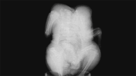
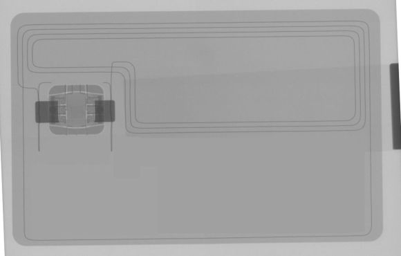
Since these RFID chips use antennas made of copper wire, he could have just started slicing his card with a knife to break the antenna — but, since he has access to a CT scanner, he thought he’d scan it to figure out where everything was.

Simply make a small notch in the edge of your card, or snip off one of the corners. This breaks the antenna and prevents power to the chip when held near a reader — though if you don’t have access to a CT scanner you might want to double-check next time you buy something!

Now there is another side to this — maybe you actually like the whole tap to pay thing, well, if you wanted to you could get a supplemental card, dissolve it in acetone, and then install the RFID chip into a finger ring for Jedi-like purchasing powers!

Converting CTs And MRIs Into Printable Objects

People get CT and MRI scans every day, and when [Oliver] needed some medical diagnostic imaging done, he was sure to ask for the files so he could turn his skull into a printable 3D object.

[Oliver] is using three different pieces of software to turn the DICOM images he received from his radiologist into a proper 3D model. The first two, Seg3D and ImageVis3D, are developed by the University of Utah Center for Integrative Biomedical Computing. Seg3D stitches all of the 2D images from an MRI or CT scan into a proper 3D format. ImageVis3D allows [Oliver] to peel off layers of his flesh, allowing him to export a file of just his skull, or a section of his entire face. The third piece of software, MeshMixer, is just a mesh editor and could easily be replaced with MeshLab or Blender.

[Oliver] still has a lot of work to do on the model of his skull – cleaning up the meshes, removing his mandible, and possibly plugging the top of his spinal column if he would ever want to print a really, really awesome mug. All the data is there, though, ready for digital manipulation before sending it off to be printed.

Continue reading “Converting CTs And MRIs Into Printable Objects”

Improving A Homebrew CT Scanner With Barium

CTscanner

[Peter] has been working on his homebrew CT scanner for a while, and it’s finally become something more than a spinning torus of plywood. He’s managed to image the inside of a few pieces of produce using an off-the-shelf radiation detector and a radioactive barium source

When we last saw [Peter]’s CT scanner, he had finished the mechanical and electronic part of the Stargate-like device, but the radioactive source was still out of reach. He had initially planned on using either cadmium 109 or barium 133. Both of these presented a few problems for the CT scanner.

The sensor [Peter] is a silicon photodiode high energy particle detector from Radiation Watch this detector was calibrated for cesium with a detection threshold of around 80keV. This just wasn’t sensitive enough to detect 22keV emissions from Cd109, but a small add-on board to the sensor can recalibrate the threshold of the sensor down to the noise floor.

Still, cadmium 109 just wasn’t giving [Peter] the results he wanted, resulting in a switch to barium 133. This was a much hotter source (but still negligible in the grand scheme of radioactivity) that allowed for a much better signal to noise ratio and shorter scans.

With a good source, [Peter] started to acquire some data on the internals of some fruit around his house. It’s still a slow process with very low resolution – the avocado in the pic above has 5mm resolution with an acquisition time of over an hour – but the whole thing works, imaging the internal structure of a bell pepper surprisingly well.

Towards A Low Cost, Desktop CT Scanner

For [Peter Jansen], the most interesting course in grad school was Advanced Brain Imaging; each class was a lecture followed by a trip to the imaging lab where grad students would take turns being holed up in a MRI machine. A few years into his doctorate, [Peter] found himself in a very opportune situation – his local hackerspace just acquired a shiny new laser cutter, he had some free time on his hands, and the dream of creating a medical imaging device was still in the back of his mind. A few weeks later, the beginnings of an open source CT scanner began to take shape.

This isn’t an MRI machine that [Peter] so fondly remembered from grad school. A good thing, that, as superconducting magnets chilled with liquid helium is a little excessive for a desktop unit. Instead, [Peter] is building a CT scanner, a device that takes multiple x-ray ‘slices’ around an axis of rotation. These slices can then be recompiled into a 3D visualization of the inside of any object.

The mechanics of the build are a Stargate-like torus with stepper motor moving back and forth inside the disk. This, combined with the rotation of the disk and moving the bed back and forth allow the imager to position itself anywhere along an object.

For the radioactive detector, [Peter] is using a CCD marketed as a high-energy particle detector by Radiation Watch. Not only does this allow for an easy interface with a microcontroller, it’s also much smaller than big, heavy photomultiplier tubes found in old CT scanners. As for the source, [Peter] is going for very low intensity sources, most likely Barium or Cadmium that will take many minutes to capture a single slice.

The machine operates just above normal background radiation, so while being extremely safe for a desktop CT scanner, it is, however, very slow. This doesn’t bother [Peter], as ‘free’ time on a CT scanner allows for some very interesting, not seen before visualizations, such as a plant growing from a seed, spreading its roots, and breaking the surface as a seedling.

[Peter] still has some work to do on his desktop CT scanner, but once the stepper motor and sensor board are complete, he should be well on his way towards scanning carrots, apples, and just about everything else around his house.

[Ben Krasnow] Builds A CT Scanner

gif

After building a homebrew x-ray backscatter imager, [Ben Krasnow] realized he had nearly all the components to build his own CT scanner, able to make a 3D model of the inside of a frozen chicken.

Basically, a CT scanner takes dozens of x-rays of an object and reassembles them with the help of fancy algorithms to allow doctors to peer inside a human body. The CT scanners you’ll find at your local hospital are monstrous devices, rotating an x-ray tube and sensor around a patient with the help of some very heavy duty electromechanical engineering. [Ben] wanted to keep his build rather small, so instead of rotating the x-ray tube and screen around an object, he simply made a stepper motor-driven lazy suzan to rotate his frozen bird.

[Ben] set a digital camera off to the side of his build and captured 45 images of a rotating chicken. After correcting for the perspective distortion, the images were thrown into 3D Slicer to create a true 3D representation of a x-rayed chicken.

Continue reading “[Ben Krasnow] Builds A CT Scanner”

Build Your Own CT Scanner

[Linas] built himself an x-ray generator for a scholarship contest. We assume this wasn’t enough of a challenge for [Linas] because after the x-ray generator was done, he used his project to model objects in 3D (Google Translate link). It’s an amazing build, leaving us feeling sorry for the guy that came in second place to the home-made CT scanner.

The theory behind a CT scanner is fairly simple – take a series of x-rays of an object around an axis of rotation. From there, it’s a fairly simple matter to digitize the x-ray images to produce a 3D model. The hard part is building the x-ray generator. [Linas] used directional x-ray tubes, a few power supplies and from what we can gather x-ray film instead of a CCD sensor. The film was scanned into a computer and reassembled to get a 3D image.

[Linas] doesn’t seem too keen on giving away the schematics for his build to any old joker on the Internet because of the high voltage and radiation components of his build. Still, it’s an amazing build.

Check out the YouTube demo of [Linas]’s CT scanner imaging an old computer mouse and a reconstruction of the same data done in MATLAB after the break.

Continue reading “Build Your Own CT Scanner”