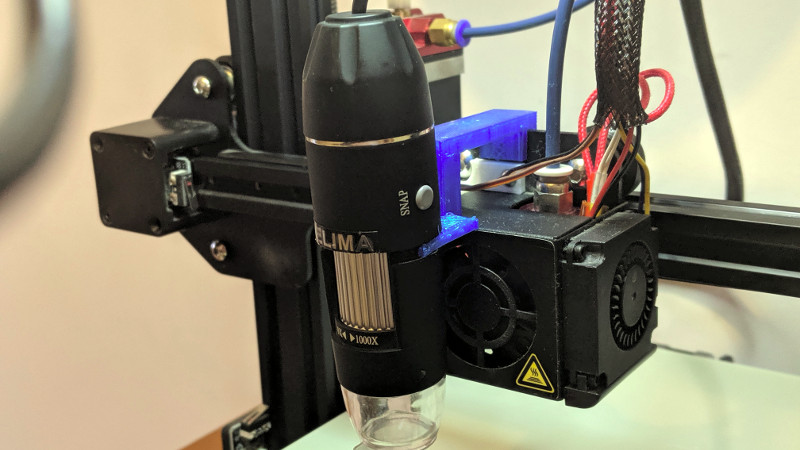
There are many ways to keep an eye on your 3D printer as it churns out the layers of your print. Most of us take a peek every now and then to ensure we’re not making plastic vermicelli, and some of us will go further with a Raspberry Pi camera or similar. [Uri Shaked] has taken this a step further, by adding a USB microscope on a custom bracket next to the hot end of his Creality Ender 3.
The bracket is not in itself anything other than a run-of-the-mill piece of 3D printing, but the interest comes in what can be done with it. The Ender 3 has a resolution of 12.5μm on X/Y axes, and 2.5μm on Z axes, meaning that the ‘scope can be positioned to within a hair’s-breadth of any minute object. Of course this achieves the primary aim of examining the integrity of 3D prints, but it also allows any object to be tracked or scanned with the microscope.
For example while examining a basil leaf, [Uri] noticed a tiny insect on its surface and was able to follow it with some hastily entered G-code. Better still, he took a video of the chase, which you can see below the break. From automated PCB quality control to artistic endeavours, we’re absolutely fascinated by the possibilities of a low-cost robotic microscope platform.
[Uri] is a perennial among Hackaday-featured projects, and has produced some excellent work over the years. Most recently we followed him through the production of an event badge.


Now just add controls for stage movement, focus and zoom and you have a whole new application for an already low cost hardware platform. Input hardware can be a low cost game controller. If restricted to the microscope functionality using the filament stepper for zoom would replace the various objectives on the typical microscope.
I guess, the real lesson here is to WASH YOUR PRODUCE
Nah it’s just more protein ;) hehe
Are you really vegan if you eat the extra protein on your produce?
No one is really vegan. They all get drunk and eat a cheeseburger now and then.
You aren’t a vegan if you breathe in air.
Ignoring the fact that insects have 6 legs, (not eight) what a great adaption of an existing technology!
Ahh.. but is your palate sufficiently discerning to be able to tell the difference between an arachnid and an insect?
I was kind if thinking the same thing, but I feel like maybe the two front “legs” are actually antennae. It’s a little hard to tell from the video, but maybe it’s an aphid?
The title of the video has a clue. ;)
He mite not get the clue ;)
I have been using a microscope (endoscope) for some time to better understand the extrusion process but bugs are cool too.
https://youtu.be/BYzClTw4kwI
I’m NOT clicking on that link until I know WHAT extrusion process you are trying to find out more about.
(fecal matter is extruded, and I have no interest in watching that!)
It’s a 3D printer. Nothing biological.
It would be interesting to combine the motion capability with some control software that will automatically track a moving target in 3 dimensions. Even if it worked in just 2 dimensions, it could be used to track swimming protists as they race around a water drop on a slide.
This is a feature I would like to implement in my own scanning microscope. You wouldn’t happen to be a programmer, would you? :)
With a stepper motor for magnification intensity control, a focus algorithm for the Z-axis and an external calibration target, or maybe an internal calibration target like a pocket comparator, seems can make an optical coordinate measuring system with. Seems better individual LED control on the LED ring light can help also for certain shapes. Neat to see!
What do you mean by optical coordinate measuring system? Like a scanner?
Before we even get to machine vision type things, how about a system to simply scan across an area and produce super high resolution microscopy images by stitching together snapshots and a google maps style interface?
It might be interesting to have a touch pad and a Raspberry-pi involved so that your finger movement would direct the X-Y motion of the scope to trail items of interest more robustly. ( and maybe a second touch pad for magnification and focus…)
Focus the microscope on the nozzle with high speed capture and you might have something useful for 3D printers.
It could also be set up to take pairs of pics to create microscopic stereoscopic images.
Has anyone thought about using an adapted microscope to more accurately tune the extruder height by analysing the filament extrusion in real-time?