We all know CERN as that cool place where physicists play with massive, superconducting rings to smash atoms and subatomic particles to uncover secrets of matter in the Universe. To achieve this aim, they need to do a ton of research in other areas, such as development of special particle detectors.
While such developments are essential to the core research needs of the Centre, they also lead to spinoff applications for the benefit of society at large. One such outcome has been the Medipix Collaborations – a family of read-out chips for particle imaging and detection that can count single photons, allowing X-rays and gamma rays to be converted to electrical signals. It may not be possible for us hackers to get our hands on these esoteric sensors, but these devices are pretty interesting and deserve a closer look. Medipix sensors work like a camera, detecting and counting each individual particle hitting the pixels when its electronic shutter is open. This enables high-resolution, high-contrast, noise hit free images – making it unique for imaging applications.
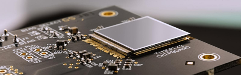
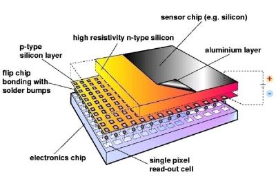
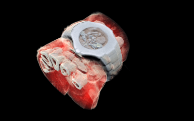 Some months back, CERN announced the first 3D color X-ray of a human made possible using the Medipix devices. The result is a high-resolution, 3D, color image of not just living structures like bones, muscular tissues and vessels, but metal objects too like the wrist watch, seen in the accompanying photograph. The Medipix sensors have been in development since the 1990’s and are presently in their 4th “generation”. Each chip consists of a top semiconducting sensor array, made from gallium arsenide or cadmium telluride. The charge collected by each pixel is transported to the CMOS ASIC electronics via “bump bonds”. The integration is vertical, with each sensing pixel connected via the bump bond to an analog section followed by a digital processing layer. Earlier versions were limited, by technology, in their tiling ability for creating larger matrices of multiple sensors. They could be abutted on three sides only, with the fourth being used for on-chip peripheral logic and wire-bond pads that permit electronic read-out. The latest Medipix4 Collaboration, still under some development, eliminates this short coming. Through-silicon-via (TSV) technology provides the possibility of reading the chips through copper-filled holes that bring the signals from the front side of the chip to its rear. All communication with the pixel matrix flows through the rear of the chip – the peripheral logic and control elements are integrated inside the pixel matrix.
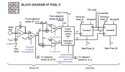
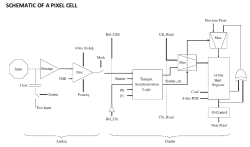
 The Analog front end consists of a pre-amplifier followed by a window discriminator which has upper and lower threshold levels. The discriminator has four bits for threshold adjustment as well as polarity sensing. This allows the capture window to be precisely set. The rest of the digital electronics – multiplexers, shift registers, shutter and logic control – helps extract the data.
Further development of the Medipix (Tech Brief, PDF) devices led to a separate version called Timepix (Tech Brief, PDF). These new devices, besides being able to count photons, are capable of two additional modes. The first mode records “Time-Over-Threshold”, providing rough analog information about the energy of the photon. It does this by counting clock pulses for the duration when the signal stays above the discrimination levels. The other mode, “Time of Arrival”, measures arrival time of the first particle to impinge on the pixel. The counters record time between a trigger and detection of radiation quanta with energy above the discrimination level, allowing time-of-flight applications in imaging.
Besides medical imaging, the devices have applications in space, material analysis, education and of course, high energy physics. Hopefully, in a few years, hackers will lay their hands on these interesting devices and we can get to know them better. At the moment, the Medipix website has some more details and data sheets if you would like to dig deeper. For an overview on the development of such single photon detectors, check out this presentation from CERN – “Single X-Ray Photon Counting Systems: Existing Systems, Systems Under Development And Future Trends” (PDF).


Interesting how they took a leaf from the human eye and compare pixels. This concept could quite easily be modified to add hardware based edge detection.
A leaf from your eye? That sounds painful.
Motes even more so.
Yes, the mote in your eye certainly looks more painful than the beam in mine.
“While such developments are essential to the core research needs of the Centre, they also lead to spinoff applications for the benefit of society at large.”
Indeed, looking at the chip I can see technology from other developments coming together. A bit of synergy as it where.
I would not be surprised if this chip (or a decendant of this chip) will be put into the first medical tricorder.
it’s crazy where we are going, just last night i was watching some hospital tv series where they are diagnosed brain aneurysm and i thought how cool would it be to have a machine to check all sort of these things, just like that we have machines to check our blood pressure we could have a small mri machine, so when my head is hurt, just to be sure, i would check my brain :) yes it sounds very funny and crazy, but who knows what the future will bring
Gee, after all that buildup and the word color, I was hoping for an imaging X ray spectrometer…after all, false color can be added to anything, using some mapping from the basic data (I do it in my own work). How about real color?
The detector is an imaging X-ray spectrometer, real colour, with the caveat that the energy range is a little limited but can be shifted using buildup material. The colour image of the wristwatch is misleading (perhaps in an effort to simplify the message to be understood). I’ve seen some results from earlier iterations of the medipix, it spits out an enormous amount of data, so much so that it maxes out the the optical fibers coming from it. These detectors are real game changers in the medical world, it’s just a bit unfortunate how expensive they are. Credit to them, they lend their test benches for research they think could lead somewhere commercially, but if you don’t have a commercial interest for them then you gotta pay, and probably can’t afford it.
I think that by “color” they mean “multifrequency x-ray”. This is: the sensor is able to specify not only the intensity of the incoming photon, but also its frequency, and since each frequency has different absortion ratios, you can create a true “color” picture, not just a picture where you paint light gray with color X, and dark gray with color Y.
The intensity of any photon is 1.0. The energy is the “color” If you get n (which is the intensity they way most people use that word), and the total is, say, 100kev, the question is, was it two of 50, a 60 and a 40, a 90 and a 10, 3 of 33.3333, and so on. If you have to keep up with individual ones, the intensity had better be pretty darn low or the data rate pretty insanely extreme as Potato says – which would be really cool if so. Doubt they’d be interested in my little radiation producing hobby (under unity fusion stuff) but then again, I have a few patrons (Not on Patreon! The real old skool type.)…I’ll ask around. Maybe they’ll lend me one for a bit, you never know.
They are imaging x-ray spectrometers with the caveat that the energy range is a little limited but can be shifted using build-up material. I had the opportunity to see an older iteration of the medipix and it spits out an enormous amount of data, even maxing out its fiber optic connection. The detectors are expensive though, to their credit they do lend out their testbenches to research that they see a commercial potential in but if they dont think you have that then you gotta pay and probably an’t afford it. Which is understandable but is still a shame as it limits the applications, for which there would be many.
Impressive, most impressive.
If they can measure individual photon energy, can they be used to build a camera that records an image while simultaneously measuring the source’s spectrum?
That’s exactly what it does, one photon at a time.
Though, actually, it’s not measuring the *source* spectrum. It’s measuring the spectrum after it passes through the object, which is the whole point.
What’s impressive about this technology is not the “color x-ray imaging” part. That part’s easy — I personally was doing it with clinical patient CT images in 1989.
What’s impressive is the combination of incremental improvements in detectors and data transmission rate.
The three key game changers this promises (but has yet to really deliver) is a) photon counting (which gives good image quality:dose efficiency), b) good photon energy discrimination (which gives good contrast:noise), and c) speed (which means decent patient throughput and lower cost). Any one of those is (relatively) easy. Getting all three at clinically useful quality for a reasonable price is mindbendingly difficult, and is what has occupied Medipix and MARS Bioimaging for well over a decade. A slow CT of a small wrist notwithstanding, it’s the technology of the future, but may always be so.
The source is a constant, therefore only needs to be measured once, whenever it is changed or re-calibrated. By subtracting the source measurement, you can learn what variances or anomalies may exist in the patient or object being analyzed, which may be useful in determining what specific condition is present.
I have been working with this technology for the last 18 years, and working directly with the Medipix / Mars Bioimaging group for the last 6 years.
The definition of COLOR:
The word COLOR is very subjective outside of the visible spectrum that the human eye can see. In the visible spectrum we can all agree on the color “Blue”. Its a specific wavelength of energy that the human eye can measure.
This detector technology is no different that the human eye, and can measure the color / wavelength of individual photons in the X-ray energy band. However, when I show you an image produced with this detector technology (or any other X-ray detector), how do we agree on the COLOR? What COLOR is an image produced with photons in the 20keV – 30keV energy band? We cant agree, because there is no corresponding color that we can agree upon. Therefore, ALL X-ray images must have their color mapped to something we can understand, it could be simple gray scale to indicate intensity, or perhaps Hounsfield units as we see in CT……. So by definition, all X-ray imaging of any kind is FALSE COLOR, because there is no such thing as TRUE COLOR, as we understand it, outside of the visible spectrum.
However, its a bit more complicated that that. Unlike all other X-ray detectors the Medipix has up to eight simultaneous energies / colors. We can use these X-ray COLOR’s to find out interesting things about what material(s) these photons have passed through (X-ray is transmission imaging, not reflective imaging like we are used too). The image of the wrist and watch does not represent the energy of the photons measured by the detector. The image is a COLOR mapped image of the materials the photons have passed through. The image represents a 3-D volume of lipid, bone, soft tissue, and metal. So to make it easy on us Humans, those different material channels, have been COLOR mapped to something we can interpret / AKA their colors in the real world. In other words, bone is a shade of white, soft tissues are a share of red, fats are a shade of yellow, and metals are a silvery gray. So the image represents a TRUE approximation of COLOR based on the materials present in the real world. However, it is still and X-ray image so……. I can now see the COLOR’s of tissues inside your body, colors that are hopefully NOT normally visible. So I am not sure how you can get any more TRUE COLOR than that.
The true value of this technology provides is the non destructive material decomposition of scanned objects. This has applications in so many fields it would be difficult to list them all. Specifically in medical imaging, this technology opens up a whole new fields of diagnostic medicine and research that were simply not possible before. By using targeted nano particle contrast agents (think Gold, I, Gd or other heavy elements) we have the perfect fusion of anatomical and molecular imaging. Think about a targeted Au nano particle that seeks out breast tumor micro calcification’s. Ask the scanner to identify gold as a material, and we can now have a 3-D image that shows us where the cancer is located on a bio chemical level. This and so much more has already been done in mice, pretty cool!
BTW: the Medipix can work with Si, GaAs, CdTe, or CdZnTe detectors. The images seen were produced using CdZnTe detectors.
Yes, color is an effective way to increase the apparent dynamic range of an image so as to more effectively visually separate image parts without degrading detail. For example, in microscopy the slides are often stained with dyes of contrasting colors to make differing areas, that may be very subtle, more obvious. Monochromatic X-ray images often have areas of importance that are similar in density appear to blend in with other areas that are very different chemically or structurally. Just increasing the contrast may improve visual separation in part of the scale, but at the expense of other parts that then appear too light or too dark for accurate analysis. By keeping the contrast lower (which widens the visible dynamic range), but using color as a differentiator, these slight differences in gradation become easier to detect.
Other differentiators may also be used, such as modulated frequencies like when combining sonograms with X-rays. Another method that is very effective and useful, especially before surgical procedures, is that of stereoscopic depth, which not only differentiates but also establishes the volumetric position of different elements in relation to others.
Combining these methodologies can be very effective in revealing differentiation and identifying edge separation and other details.