Of the body’s organs, the lungs are among the trickiest to take a biopsy and treat cancer in, both due to how important they are, as well as due to their inaccessibility. The total respiratory surface within the average human lungs is about 50 to 75 square meters. Maneuvering any kind of instrument down the endless passages to reach a suspicious area, or a cancerous region to treat is nearly impossible. This has so far left much of the lungs inaccessible.
The standard of care for lung cancer is generally surgical: remove parts of the lung tissue. However, a proposed new method using magnetic tentacles may soon provide a more gentle approach, as described in Nature Engineering Communications by Giovanni Pittiglio and colleagues (press release).
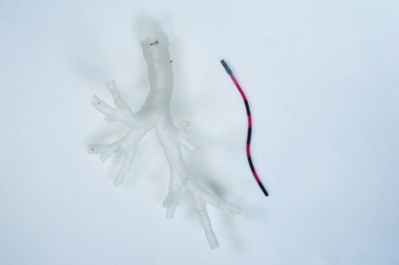
The tentacles are made out of a silicone substrate with embedded magnets that allow for it to be steered using external magnetic sources. With an embedded laser fiber, the head of the tentacle can be guided to the target area, and the cancerous tissue sublimated using an external laser source. In experiments on cadavers with this system, the researchers found that they could enter 37% deeper into the lungs than with standard equipment. The procedure was also completed with less tissue displacement.
Considering the high fatality rate of lung cancers, the researchers hope that this approach could soon be turned into a viable therapy, as well as for other medical conditions where a gentle tentacle slithering into the patient’s body could effect treatments previously considered to be impossible.
Heading image: Close-up of a magnetic tentacle robot next to a phantom bronchiole (Credit: University of Leeds)


This is quite clever and obvious after someone else thought of it.
“could affect treatments previously considered to be impossible.”
Effect, not affect.
That would be my bad in the edit. Maya had it correct.
“cancerous tissue sublimated ” is a pretty neat trick. I mean, after you heat the tissue with the laser enough to first pyrolyse it at 600 C, the remaining carbon will sublimate around 3600 C. Or catch fire, whichever comes first.
The word is actually “ablated”
can i nitpick/play devils advocate here :P
laser ablation is more or less like a microscopic explosion ( unless you turn the power down and increase time but i don’t think making “lung roast” is the point here) so basically it turns cells into
micro-particles which although not gas per se is close enough. so its maybe not correct in the technical sense but i would argue it close enough in the literal sense.
Putting smoke into the lungs doesn’t seem terribly healthy. It might give someone lung cancer.
The original authors use “ablate”, as do practitioners of similar art. In context it does not mean “explode” or “turn into smoke”; it’s taken to mean “destroy” or “remove”. “Cook” works too, but patients don’t like to hear that one.
“The standard of care for lung cancer is generally surgical: remove parts of the lung tissue. However, a proposed new method …”
“… the researchers found that they could enter 37% deeper into the lungs than with standard equipment. ”
The article doesn’t seem to reflect what the standard is very well unless the standard equipment is for imaging/biopsy only.
With that, I’ll relate a story from a colleague who was involved in U.S. regulatory approval for a similar device: At some point the FDA-CDRH kept requesting more and more data about it, requiring animal (rat) studies etc. which delayed approval and caused the expected amount of stress and expense. At some point they sent in the results for a similar study done on the device in Canada. The FDA’s response was (allegedly) “Oh, okay, we don’t need data from rats if you have it from Canadians.”
It sounds just like the method we use to thread internal hydraulic brake hose through a bicycle frame.
A magnetic worm and a magnet.
This article is a new low. Posh and HaD editors: pretty please stop regurgitating the outlandish press release claims of research papers that claim to revolutionize a field. It is a bad look. It is dishonest. It is bad reporting. If you are going to report medical stuff, at least have an RN take a look at it first or literally anyone that works in a medical field.
The lungs are absolutely NOT the trickiest to take a biopsy. There are many, many routine ways of doing it. Starting with a bronchoscopy which is performed (routinely) with either a rigid bronchoscope or a flexible fiberoptic bronchoscope. A rigid bronch can do washings, then that liquid (gets down reallllly far… its a liquid!!) is spun down and the cytology examined by the pathologist. Flex fiberoptic scopes: The smallest are about the diameter of a piece of angel hair pasta and can easily get down “real far” too. If that isn’t good enough, you can easily (routinely) do a percutaneous biopsy that is CT guided. A needle is guided to the tissue site under CT guidance.
Cancer treatment for lung cancers is a huge spectrum ranging from chemotherapy, rad-onc approaches (zero invasive gamma knife for example), to, yes, surgical excision.
Ablating cancer cells by any means, by the way, has been problematic in other cancers. Because you don’t necessarily kill every single cell then all you are doing is spreading it around.
The research article addresses most of those points you raised. It might be worth it to read it first before commenting.