Mapping A Fruit Fly’s Brain With Crowdsourced Research

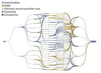
Example of a graph representation of one identified network with connections coded by neurotransmitter types. (Credit: Amy Sterling, Murthy and Seung Labs, Princeton University)

Compared to the human brain, a fruit fly (Drosophila melanogaster) brain is positively miniscule, not only in sheer volume, but also with a mere 140,000 or so neurons and 50 million synapses. Despite this relative simplicity, figuring out how the brain of such a tiny fly works is still an ongoing process. Recently a big leap forward was made thanks to crowdsourced research, resulting in the FlyWire connectome map. Starting with high-resolution electron microscope data, the connections between the individual neurons (the connectome) was painstakingly pieced together, also using computer algorithms, but with validation by a large group of human volunteers using a game-like platform called EyeWire to perform said validation.

This work also includes identifying cell types, with over 8,000 different cell types identified. Within the full connectome subcircuits were identified, as part of an effort to create an ‘effectome’, i.e. a functional model of the physical circuits. With the finished adult female fruit fly connectome in hand, groups of researchers can now use it to make predictions and put these circuits alongside experimental contexts to connect activity in specific parts of the connectome to specific behavior of these flies.

Perhaps most interesting is how creating a game-like environment made the tedious work of reverse-engineering the brain wiring into something that the average person could help with, drastically cutting back the time required to create this connectome. Perhaps that crowdsourced research can also help with the ongoing process to map the human brain, even if that ups the scale of the dataset by many factors. Until we learn more, at this point even comprehending a fruit fly’s brain may conceivably give us many hints which could speed up understanding the human brain.

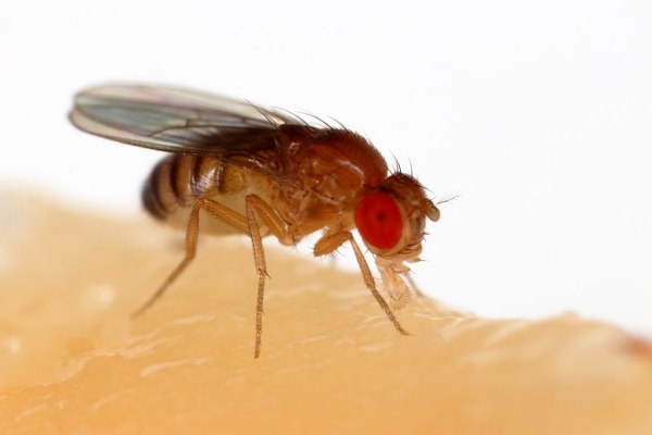
Featured image: “Drosophila Melanogaster Proboscis” by [Sanjay Acharya]

FlyBrainLab: Google Earth But For A Drosophila Fly’s Brain

In biology there are a couple of truly crucial model animals and insects. Not that they’re particularly good students, or pick up their own trash, but in the sense that they have become standard model organisms for research. Aside from genetic research, the FlyEM project seeks to fully map a little fly’s brain’s neural connections. This common fly, called drosophila melanogaster (or ‘lesser fruit fly’) has been the subject of a lot of genetic studies, but this study of its brain structure may provide insights in how our brain works as well.

Based on electron microscope images of thin slices of a drosophila brain, the three-dimensional structure of this tiny brain is reconstructed to not only determine the location of each neuron, but also their connections with other neurons. We know that about two-thirds of their brain is dedicated to processing the visual information from their relatively advanced compound eyes, but a lot is still unknown about how this is done, or how the brain’s structure develops.

If it’s always been your dream to tinker with a little fruit fly’s brain, you can do so yourself using the open source FlyBrainLab tool provided, along with the freely available data sources. This tool does not just allow one to visualize the drosophila brain in great detail, but also to create executable circuits and study their functionality. With neurobiology still a largely unexplored territory, this makes for an amazing tool to make this research accessible to anyone.

(Thanks for the tip, [Hernandi Krammes])