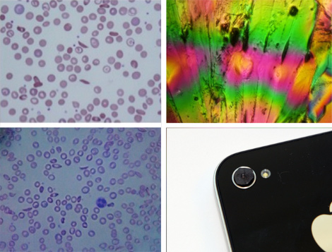
Medical-grade microscope photography for $20 might be a game changer in areas where medical services are unavailable. This particular hack uses an iPhone’s 2 megapixel camera, as well as a tiny glass marble, to magnify a sample to about 350 times its actual size. The two images seen on the left are red blood cells photographed with the improvised microscope. The main issue with this magnification method is a very thin plane of focus that is overcome with processing in software.
This makes us think of the microscope hack that shined a laser through a droplet of water, to project the image on a wall. The concept was later refined to work with samples on glass slides. There are a couple of distinct advantages to using this cellphone-based method. First, the sample can be seen with its true colors. Second, you not only magnify the sample, but you have a digitized image already on a device that connects to the Internet. If you’re trying to make a medical diagnosis this can easily be sent to a qualified professional for analysis.
The team that came up with this technique also figured out how to build a cellphone-based spectrometer for just a few bucks. The image in the upper right is the result of that hack. Both have a step-by-step build guide on the page linked at the top. The microscope is just a glass bead in a piece of rubber, as seen here. The spectrometer is a bit more involved.
[Thanks Fabien]


So how do they take multiple images to do the focus stacking? I’d think handholding the phone would not be suitable.
Thus the issue of lack of stabilization, digital or optical, on phone cameras rises again.
iphones have more than enough power to do base cell simulation, but I’d hate to be the one paying the devs on that. Ham ‘n’ Egg devs can’t touch this so indian software firms and teenagers are out of the question..
Gorillapod.
Combine the two and you’ve got an invaluable in-the-field microscope for poor and developing areas (aussie outback, africa etc).
They very tight on memory. Most devs have to employ wacky tricks to get big apps to run.
Now, a cheap webcam and laptop would be far more practical; as it stands this article is another thinly veiled ad for a $500 phone.
Reading about this microscope, at no point do its devs say that an iPhone is required, nor that the software runs on a phone. Their pictures of various microscopes show a range of phones.
Yeah any phone could do it, it’s the marble that does the magnification.
Most phones have a DSP and GPU to go along with the main instruction logic. This is just on the app processor too, the baseband could also be used through hacky code.
I doubt it’d be needed but storing pages in a file would work for any memory issues.
I’ve had good results with just holding/taping a handheld microscope from the shack to the lens of an old VGA camera – first you focus the phone and then dial the focus in on the microscope. Here’s the cheapo I used http://www.radioshack.com/product/index.jsp?productId=2179604 $12 and 60-100x zoom. Probably won’t see any blood cells, though.
Good enough for removing slivers?
Could the processing power of the phone be used to automatic run test and compare cells.
I have 0% knowledge of medicine, so i might be blowing smoke
You can do this too with a laser. Just replace the lens (in [1]) with a pinhole.
If your laser is powerful enough, you can even do a reflection setup instead of transmission (where you see the shadows of the atoms):
     (projection)

(laser)  \  /

   \      \/

    \   (lens)

     \  ~~

      \~~

    (sample)
If you want to further improve results, grab a lens with a small focal length and focus the light coming from the sample into a pinhole [2]. This will allow the first order frequencies to pass and reduce aliasing (visible in the video):
    (projection)

        \  /

         \/

     (pinhole)

(laser)  /\

  \     /  \

   \   (lens)

    \  ~~

     \~~

  (sample)
Hope my ascii drawings preserve their format…
[1] http://hackaday.com/2011/03/02/a-different-take-on-a-laser-projection-microscope/
[2] http://en.wikipedia.org/wiki/Spatial_filter
You can do this too with a laser. Just replace the lens (in [1]) with a pinhole.
If your laser is powerful enough, you can even do a reflection setup instead of transmission (where you see the shadows of the atoms):
(projection)
(laser) \ /
\ \/
\ (lens)
\ ~~
\~~
(sample)
If you want to further improve results, grab a lens with a small focal length and focus the light coming from the sample into a pinhole [2]. This will allow the first order frequencies to pass and reduce aliasing (visible in the video):
(projection)
\ /
\/
(pinhole)
(laser) /\
\ / \
\ (lens)
\ ~~
\~~
(sample)
HaD: Please remove my last comment, it was a failed attempt on ascii drawing..
[1] http://hackaday.com/2011/03/02/a-different-take-on-a-laser-projection-microscope/
[2] http://en.wikipedia.org/wiki/Spatial_filter
You can do this too with a laser. Just replace the lens (in [1]) with a pinhole.
If your laser is powerful enough, you can even do a reflection setup instead of transmission (where you see the shadows of the atoms):
http://pastebin.com/mmyXvRUE
If you want to further improve results, grab a lens with a small focal length and focus the light coming from the sample into a pinhole [2]. This will allow the first order frequencies to pass and reduce aliasing (visible in the video):
http://pastebin.com/x5L6nQU3
HaD: Please remove my last comment, it was a failed attempt on ascii drawing..
PS: A preview button here would be really nice..
[1] http://hackaday.com/2011/03/02/a-different-take-on-a-laser-projection-microscope/
[2] http://en.wikipedia.org/wiki/Spatial_filter
Very nice indeed! Since there is no depth of field needed it certainly has possibilities. One part that I seem to have missed is that there is post image software manipulation. Did I miss something?
This is basically a ‘van Leeuwenhoek’ style microscope with a cell phone camera serving the role of the human eye. They could have saved twenty dollars and called this a ‘free’ microscope by making the lens the same way Anton van Leeuwenhoek did in the sixteen hundreds. He took a piece of glass, heated it and stretched out a fiber, then melted the end of the fiber until it formed a spherical drop. If they’re planning on examining blood smears, they’ve already got some microscope slides. The glass in microscope slides is much better optical glass than the soda lime glass van Leeuwenhoek used; with a microscope slide and a blowtorch, you could make hundreds of lenses, for little more cost than the time spent to do it. This allows a more durable and stable mount as well, as the lens has a ‘tail’ attached to it. You can make an aperture in a disk of thin metal, then glue the tail of the lens to the metal with nail polish, resin, shellac, or even wax.
its funny that they call Leeuwenhoek microscope a $10K lab equipment
The top right image is not a spectrophotometric image. It appears to be a crystal viewed with the microscope with a polarizer in the image path.
But, couldn’t it be processed (e.g. The GIMP) to extract wavelengths (colors) and possibly intensity (amount of amount of each color)?
Doesn’t that iPhone’s camera have an 8- (or 5-)megapixel sensor? Is this just usual Szczys or am I missing something here? Is there a de-Bayer-ization factor being applied?
Cool, let me know where I can buy one of these $20 iPhones.
*shone
*Neither* the top left or right images are from the cellphone camera. They’re from an actual lab microscope.
The *bottom* images are both from the cellphone camera, for comparison. The left images are sickle-cell anemia blood cells, and the right images are of a crystal.
How much image processing is really necessary? The image in a Leeuwenhoek microscope is fairly clear in the center, so why not just use that portion.
Very clever!
Has anyone thought of using a PS3 BRIR diode as a light source for multiwavelength fluorescence microscope a la Rife?
:-)
Great where access to medical equipment is scarce, but wireless net access is plentiful?
Where might this be?
3rd world countries…
It’s much cheaper to put up cell towers in major cities (or to attach cell antennas to existing structures) than to run POTS or Fiber through the streets. (Where the locals would steal copper wires for recycling anyway). So many 3rd world cities adopted cellular technology years ago. Besides, it’s much easier for 3rd world dictators to monitor the phone calls and movements of their enemies with cellular technology. But they don’t give a damn about health care for their citizens. If they need medical care for themselves they can often fly to Europe or the U.S. for it.
Ah, thanks!
I just didnt make the connection there. I keep forgetting how cheap this stuff is now, and how much cash flow they’ve had recently.
I was in telecom up until the dot-com crash, that’s why I still assume they had limited cell with no net access. I have a tech stereotype that’s stuck in 2001. Cant believe it’s been ten years already…whew…
We were still all supposed to have fiberoptic feeding a 4-home pedestal by 2001 (predicted in 96-97 and planned for by Ameritech, SBC, AT&T. I was in all their meetings as a subcontractor). Back then, nobody knew what the bandwidth was going to be used for anyways.
Now I just wonder where the $20 figure comes from.
Africa. Cell towers=Net access.
The spectrometer they built can hardly be called a spectrometer. All color cameras have IR blocking filters installed in them, and the cut-off characteristics of these filters will affect the data greatly (especially in the UV and NIR range). A grayscale camera is more suited for the job.
Not quite microscopic but had some fun with a lens from an old cd-rom and SE w810 cellphone. http://www.priithansen.com/makro.png
great for me, i used a 45.00 dollar Ematic music player and a cd-rom lens not quite the 350 x power but works good for viewing micro boards, and i added an old security cam to my work table with an old camera lens on the camera. got enough room to work under the camera. hooked it to a moniter no more magnifing glass, great for working on small items.