[the_digital_dentist] had a CT Scan done back in 2007 for treatment using orthodontics. Some how, he managed to get a copy of the CT Scan data from the lab, and has been playing around with it lately.
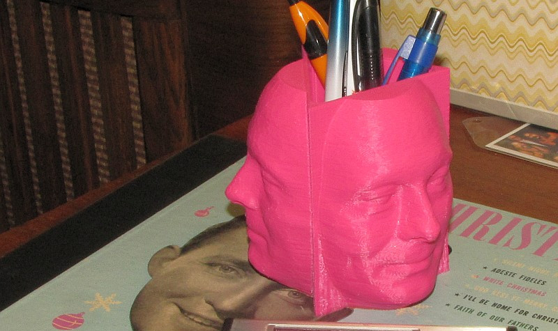
Since he has a 3D printer, the obvious end goal was to print his face using some of the data extracted from the CT Scan. This required a lot of manipulation to get it to the finished model you see above. He used an open source software called DeVIDE to process the data and export the STL. Not much information on this is given on his site, but in our research we managed to find another video documenting the process in DeVIDE on extracting the STL model from DICOM CT scan data.
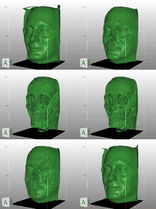 Unfortunately, the STL is far from being ready to print after being extracted; there is a lot of extraneous data that needs to be cleaned up. He used mesh editing software to help blow away the unnecessary details. We don’t know for sure what software [the_digital_dentist] used, but MeshLab is a good one.
After that, it was just a matter of printing the STL file. But the really cool thing about using data from CT scans is the amount of detail it captures… Stick around after the break to see an animated GIF demonstrating this.
Anyone want to print a copy of their own skull? It’d look great with a plating of Adamantanium…












A case for 3D metal printing if I ever saw one.
Speaking of metal printing, I’d *love* a paperweight of my own skull.
A CT scan just to print a face? Yes it was incidentally done but use a 3d scanner!
If you’ve got the CT scan done already…
Creepy.. M
“Some how, he managed to get a copy of the CT scan data…”
Just asking the technician who is doing the scan gives very good results, in my experience. I’ve received raw data for MRIs, CTs, ultrasound and endoscopy just by asking – though you sometimes get asked for £10 to cover them burning you a CD.
For processing the data, I’ve had good results with OsiriX (an OSX DICOM reader/renderer) but as the article alludes to, the hardest work is in segmenting the data afterwards – the DICOM is effectively just a series of 2D image ‘slices’ with a known thickness and each pixel’s value representing a density. Deciding what ranges of densities to filter in and out of your output polysurface is a real pain and none of the automatic tools work very well. That said, it looks great when you get it right :-)
Any diagnostic scan is *your data*. Whether is an X-ray, CT or MRI. All you have to do is ask for it on a disc in DICOM format. The imaging center/hospital *MUST* give it to you under the HIPAA law. It’s your data. The most they could do is charge you for the CD, and any hospital/imaging center that would do that would quickly go out of business.
Already had a diagnostic procedure done within the last few years? It’s not too late. Go ask for it.
For any Swedes out there you can just ask for the raw data and they’ll give it to you for a 100kr (~15USD) fee. I have a few scans of myself from when I had trouble with a kidney stone (*drinks some water as he types it*). My plan was to print out a full size copy of my pelvis and part of the spine but I found out I truly suck at cleaning up “broken” STLs. That and there was a bit of missing detail around the tail bone.
It’s really fun to play around with though. You can chose to look at only bone, only muscle, fatty tissue and stuff :)
Genitalia too?
Ooh! An HP calculator! Which one?
A 15C. The man has taste. I’ve had mine since the mid 80’s and it still works perfectly. I use it all the time.
Almost thought it was a 16C, which is what I have laying around in my office.. but it’s not.
Jealous. I want one but they cost serious $$ on eBay. I wish HP would reissue it, or at least sell something like the 35S but with hex mode.
There a credit-card sized clone available.
Check out: http://www.swissmicros.com/
I initially was hoping it was a 3D printed model of a HP1?C, and then I noticed the bright pink penholder.
Not that a functional 15C isn’t better, but.
I am the originator of this object. I’ve put more details of how the CT scan data was processed on the Thingiverse page here: http://www.thingiverse.com/thing:203856
The youtube video showing how to use DeVide was extremely helpful.
Next up: the same cup with 1 face, 1 partial face/bones exposed, 1 bones only. It will end up on Thingiverse after I finish a few printer mods/upgrades.
The CT scan was done in 2007 for orthodontics treatment planning while I was in dental school. I handed the radiology tech a blank DVD and he saved the files to it for me. Even then I anticipated this use for it. Anyone who wants to play with the actual CT scan data can download my file here: http://www.rehorst.com/mrehorst/CT%20Scan%20DiCom3.rar
It’s about 50 MB and consists of 500+ individual images.
If you use the file to print anything, please leave a comment on the Thingiverse page.
Have fun!
A CT scan of the entire head just for orthodontic use? Seems like a bit of unnecessary radiation exposure.
I’ll beg my orthodontist to give it to me :-)
Personal jelly moulds!
Actual URL for the blog post is: http://milwaukeemakerspace.org/2013/12/further-adventures-in-ct-scan-3d-printing/
Why his face both sides? Back should be his skull.
To echo some of the posters above: CT scans I’ve encountered (dental cone beam and some other variants of the same tech) save files as a series of slices of (what looks like) bitmap images. Stack the slices on top of one another and you have a greyscale cube where the value of a pixel is the density of the material at that pixel.
All the scans I’ve encountered come on a CD bundled with some reading software that does the math and applies some filters so you can do stuff like isolate the dense bone from the not-so-dense surrounding tissue. Like playing with levels in photoshop and using the smart select tool on the result. Cool stuff.
I had a CT scan done a couple of years ago, and I got ahold of the DICOM data (asked the technician for it), but when I try to open it in OsiriX and Slicer, it shows up distorted, kind of squashed-looking. Does anyone know why this may be happening or how to fix this?