I loved my science courses when I was in Junior High School; we leaned to make batteries, how molecules combine to form the world we see around us, and basically I got a picture of where we stood in the scheme of things, though Quarks had yet to be discovered at the time.
In talking with my son I found out that there wasn’t much budget for Science learning materials in his school system like we had back in my day, he had done very little practical hands-on experiments that I remember so fondly. One of those experiments was to look and draw the stages of mitosis as seen under a Microscope. This was amazing to me back in the day, and cemented the wonder of seeing cell division into my memory to this day, much like when I saw the shadow of one of Jupiter’s moons with my own eyes!
Now I have to stop and tell you that I am not normal, or at least was not considered to be a typical young’un growing up near a river in rural Indiana in the 60’s. I had my own microscope; it quite simply was my pride and joy. I had gotten it while I was in the first or second grade as a present and I loved the thing. It was just horrible to use in its later years as lens displaced, the focus rack became looser if that was possible, and dirt accumulated on the internal lens; and yet I loved it and still have it to this day! As I write this, I realize that it’s the oldest thing that I own. (that and the book that came with it).
Today we have better tools and they’re pretty easy to come by. I want to encourage you to do some science with them. (Don’t just look at your solder joints!) Check out the video about seeing mitosis of onion cells through the microscope, then join me below for more on the topic!
What is Mitosis?
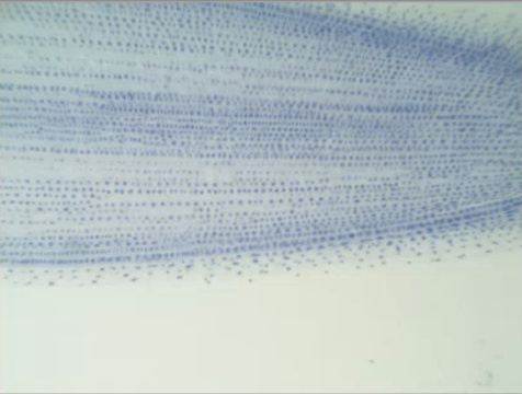
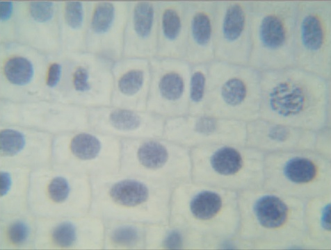
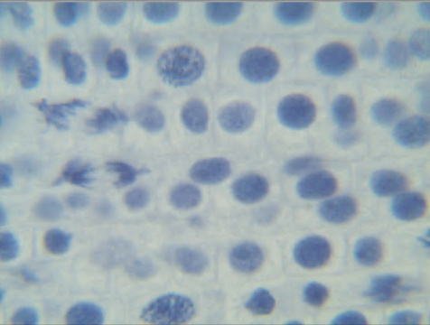
The microscope is an excellent tool of Biology and a great place to start is by observing mitosis as it happens. Mitosis is where cells divide to make more cells so that tissue may grow. As it turns out, the tips of fresh roots on onions and garlic have rapid tissue growth in a concentrated area, making the viewing of the various stages of cell division worthwhile. In preparation for this article I started growing a shallot and an onion in a class of water and waiting for the tips to spout.
mi·to·sis /mīˈtōsəs/ noun BIOLOGY
- a type of cell division that results in two daughter cells each having the same number and kind of chromosomes as the parent nucleus, typical of ordinary tissue growth.
I can think of no better example of being able to see one of the complex miracles that make life possible than to look at cell division with one’s own eyes.
Scope with a Camera
My son owns a ‘scope that is much better than my old tiny clunker, yet he got it for the same price as what mine cost back in the day relatively speaking. What’s more is that I can replace one of his eyepieces with a camera and share what I see with others — you in this case. This is something I would not have ever imagined being able to do “back in my day”.
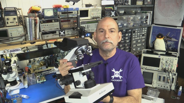 I actually have several microscopes in my hardware lab, two are dedicated to assisting with surface mount assembly, but my son’s includes up to 2000x magnification! That what I used to make these picture.
What We’re Looking For
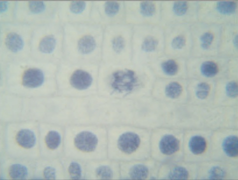
Life based on cell division has the ability to make new copies of vital tissue cells starting with the ability of DNA to divide and replicate. As the process continues, whole chromosomes replicate by division with two new cells forming in place of the original one. Different organisms have differing numbers of chromosome by default, the onion under the scope today has 16 chromosomes arranged in 8 pairs, by default compared to the 23 pairs of a human. The number of chromosomes doesn’t necessarily imply the complexity of the organism, dogs have 78 chromosomes for example, compared to humans’ 46.
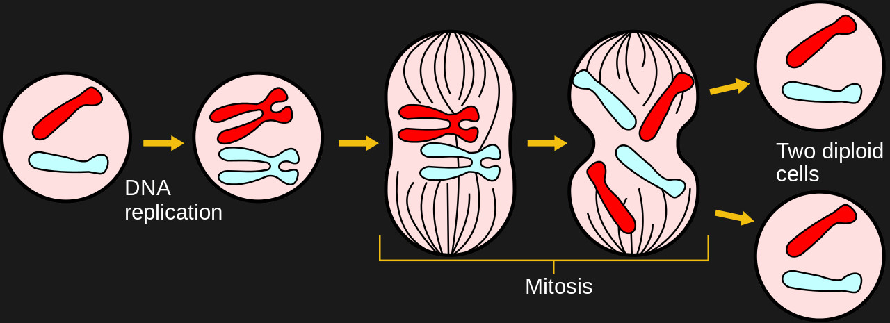
Phases
Prophase or the “before” phase. The cell still looks very similar to a non-replicating cell even though things are starting to happen.
Prometaphase is where the walls of the nucleus break apart and the act of cell division takes over the whole cell. This is where it really starts to be visible using this level of microscope.
Metaphase or “next to” and true to its name the chromosomes line up side by side near the center of cell as various forces pull in opposite directions. This looks cool when seen in real life.
Anaphase or “after” is when it starts to look like as new twin cells.
Telophase or “end”. ‘nuff said.
Sliding Away
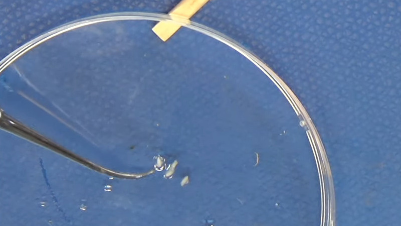
To prepare the onion tissue for observation I first soaked it in warm hydrochloric acid. If you think that that sounds like I am digesting the tissue much like our own stomachs do, you would be correct. The cells cease activity, sometimes called “fixing”. A bunch of the matter exterior to the cell walls is digested or softened, allowing us to concentrate on the contents of the cells. It also makes it easier for dye to get into the cell and to mash the tissue thin enough to see one layer of cells.
I meant to use a Feulgen stain and thought I had some. I didn’t. I ended up using Methylene Blue, an old standby and was the stain I used originally back when I was young. The slides weren’t quite as clear as I would have liked but my son still got the experience of making his own slides.
Smashing Roots
The next step is to carefully smash the softened root tissue as flat as possible in an effort to get as close to one layer of cells. Usually I break the slide cover doing this, occasionally resulting in my finger bleeding all over the slide, but today it goes almost perfectly.
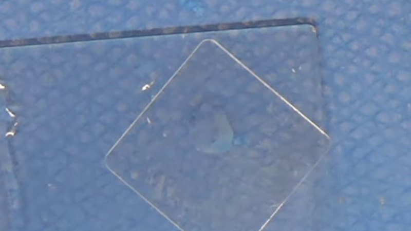
With that said, there is only so much flattening the “mash method” and part of the experience in looking through the eyepiece is continuous adjustment of the focus as the subject matter still has an amount of three dimensional aspect to it.
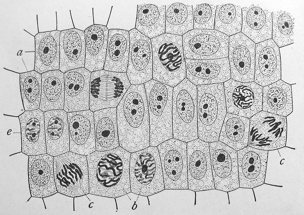
As can be seen in the images below, we caught all of the major phases of mitosis, my onions have been sacrificed for a worthy cause.
Conclusion
I loved my microscope, and still do, it represented my ability to study and learn on my own and yet see way more than I could than I could without it. It also allowed me to focus my curiosity in a hidden realm and was a early gateway in my quest for science when I was young.


FYI,
Cancer radiation therapy targets cell in mitosis, that is when the DNA of cancer cells are most vulnerable to mutation, (mutated DNA/RNA are more likely to kill a cell as opposed as making it cancerous).[citation needed]
And since “a” definition of cancer is “cells reproducing faster than normal”, the numbers of cancer cells in mitosis is high.
So, the cancerous cells are affected more than normal cells by radiation.
But, because not all cells are in mitosis at the same time, multiple radiation treatments are often given to hit cells that were not in mitosis during previous treatments.
As an aside, antioxidants prevent cancer by the suppression of oxidants which damage cell DNA (especially during mitosis).
A collateral cancer treatment is to cut back on the consumption of antioxidants and increase the consumption of oxidants, therefore increasing the damage to the cancerous cells.
Nice article
wow that volume on the end!
Heh, yeah I get a little rusty between vids.
I think He meant the melody at the end with the full volume square wave
Someone watched it through to the end???!!! :)
@Bil Herd
Is that an Omax brand model?
Omax microscopes are inexpensive, and the Achromatic objective lens tend to be fairly good. Indeed these often include a stage-position lock on the coarse focus adjustment too. ;-)
I “upcycled” a few units for a project recently, and was amazed at what $70 (new) buys these days. Two issues you may encounter is Omax prism mirror surfaces will tend to pit over time, and the low-end models often get internal dust contamination fairly easily.
Another easy project is setting up a miniature Spirolina fed aquarium for Artemia, Triops cancriformis, or Streptocephalus, Additionally, intuitively understanding Algae-bacteria interactions in aquariums will likely become a rather practical life skill in the near future.
I have three AmScopes in the lab…. I only need two but still have three as I like scopes.
I bought a ~$200 AmScope over a year ago, in addition to using it on SMD circuits, I told the wife it would be good at removing tiny slivers!
B^)
So Bil,
what is that thing you were holding in your hand at the end of the video?
It looked like the numeric keypad for my laptop without the USB cable.
Thats how I control the video, it is a wireless numeric keypad.
The easiest way to get a nice thin slither of onion cells isn’t to mash a blob of them flat.
Skip the acid. Instead of getting cells from the layers, use the thin membrane between the layers
It will peal away nicely and it’s already only a couple of cells thick. You can basically get away with simply viewing it as is or using a light stain. The good thing about onion cells is they are relatively large. You can always try your own cheek cells if you’re feeling adventurous.
I thought about showing a home made microtome. :)
Until I gggggooooooogggggled it,
I thought a microtome was a very tiny book!
(Possibly made by stacking microdots)
B^)
In 5th grade, i saved and bought a 6″ tall metal microscope for something like $12 (back in 1962, that was a LOT of money and would be over $100 today). It was small, but well made. From Japan I’m sure. I think it had 150, 300, & 600 magnification via a turret, two ways of illuminating the slide: mirror and 2 AA cell with one of those penlight bulbs with a lens to focus the light. Anyway, one particularly hot summer night (we had no A/C or even a fan), I couldn’t sleep and there was a bright, full moon shining through my bedroom window. I thought it might be bright enough to view a slide on my new microscope, so I planted the microscope on the window sill, but I saw dark “spots” that I was sure shouldn’t be there since they were stationary even when I moved the slide.. So I pulled the slide out and adjusted the scope’s up/down (focus) and found that the (dark) spots was a reflection of one of the moon’s craters in the mirror! I wasn’t real clear, since it was bouncing off the fairly distorted mirror surface (I didn’t know the difference between first surface vs. standard mirrors, let alone the importance of “optically flat” surfaces). I played some more, moving the microscope’s position to see if I could see other parts of the moon — which I did see, but it was hard as the magnification magnified my movements. I soon tired enough to fall asleep and that was the end of that adventure.
I remember learning the foibles of my early scope as I got better at using it…. but I still loved that thing.
His name is Right There!
Yeah it was wasn’t it. There is always something I would redo in every video.
Excellent concern intro… scary actual after I learned more about law of which isn’t even taught from kindergarten up as needs to be so time is better spent on valid arguments if any… and great in general video!
Reading into interferometry lately and wanting to make a interferometer spectrometer along with the dispersive (diffraction grating) webcam iterations… gets me thinking can use maybe some parts salvaged to make a projector to observe the target on a wall to better see detail?
Also, I’m wondering if can come up with a open source FTIR microscope spectrometer, Hyperspectral Imaging and/or other maybe something more suitable for needs… rapid bacterial detection for counting or microbe effusia… maybe or biopsy chemical testing?
Microscopes are awesome to see at the University Surplus stores back prior to the COVID closings. When I was looking to get a stereomicroscope for SMD soldering and in general inspection work, I struggled not to invest in a microscope for biological use on the farm. Interestingly one, that was way cost effective, appeared at a Goodwill Store and I invested in.
Makes me think that a fun handy tool might be a hacked, 3D printed maybe or machined, open source… (I had to look online since I was only thinking slicer)… Microtome .
https://en.wikipedia.org/wiki/Microtome
Why would you need a dispersive element with an interferometer? A few years ago there was an article here about an FT spectrometer in the visible range.
https://hackaday.com/2015/04/22/a-diy-fourier-transform-spectrometer/
For IR the problem will most likely be the beam splitter, the longer movement of the interferometer and the detector material. An FT microscope is quite simple, if you have acess to an FTIR spectrometer: just scan the sample. A two-dimensional IR detector, however is quite expensive.
@MS, et.al. – “Why would you need a dispersive element with an interferometer?”
I don’t know, two separate spectroscopy projects. Now you got me thinking something RAMAN or something??? :-|)
On the topic of dispersive instruments… I’m planning to use the actuator arm of a HDD to scan using a blu-ray disc… or that is still a project on my mind since the plan isn’t on paper yet… well now is sort of. Thinking the HDD body will be a neat small optical table too… for use also with a interferometer project maybe where I’m hoping to be able to use old CD/DVD/Blu-Ray optical components. Sam Zaloof recently made an awesome video on his interferometer project and I wonder how even more cost effective that system can be open source scaled down in price point to.
That get’s me thinking about this system if you’ve not seen worth sharing also:
https://www.youtube.com/watch?v=5bqujaldaCQ
On the topic of the FT instrument, thanks for the link which got me searching for more Hackaday articles. Haven’t read that article. Have the following though: https://hackaday.com/?s=hyperspectral+imaging
Didn’t see any articles on RBD or similar methods. I know micro was apprehensive of that and my confidence in using just TLC to detect certain pathogens based on their effusia and effluent… then further analyzing the specific section of the plate… or validating the methods based on the rigorous developmental analysis. Back in the days of advocating the back room low key IMS-IMS cleaning validation methods just prior to me advocating using way better the GC-SAW Z-Nose and e-notebooks with lab automation… the later two didn’t help my career at all… then add the coordinate measuring system and wow… I’m still here.
Anywho, for society in general, I’m sure there are cost effective ways to better experiment, research and mainly diagnose.
Was just checking back to post the link I randomly (I guess) just found suggested after reading two IEEE articles “Build a Long-Distance Data Network Using Ham Radio” that was suggested after reading “The Uncertain Future of Ham Radio”:
https://spectrum.ieee.org/geek-life/hands-on/build-a-sophisticated-microscope-using-lego-3d-printing-arduinos-and-a-raspberry-pi
Wow! Awesome FT article with reference and I only cursory reviewed so far! Really detailed… sweet!
Meetosis is a disease of too many meetings. It is reunionite in french.
Mee tooo I agree. >:)
“The meetings will continue until productivity improves!”
B^)
“Look at me toes, Sis!”
Loved this video. Thanks!!