Quick And Dirty Microscope Motion Control For Focus Stacking

If you’ve spent much time looking through a microscope, you know that their narrow depth of field can be a bit challenging to deal with. Most microscopes are designed to only have a very thin slice of the specimen in focus, so looking at anything above or below that plane requires a focus adjustment. It’s tedious and fussy, and that makes it a perfect target for automation.

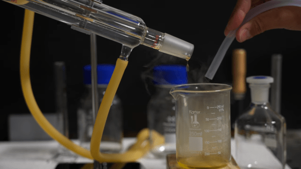
The goal behind [ItMightBeWorse]’s microscope mods is “focus stacking,” a technique where multiple images of the same sample taken at different focal planes can be stitched together so that everything appears to be in focus. Rather than twist knobs and take pictures manually, he built a simpler Arduino-based rig to do the job for him. Focus control is through a small stepper motor connected to the fine focus knob of the scope, while the DSLR camera shutter is triggered through a simple relay board. There’s also lighting control, with an RGB LED ring light that can change both the light level on the sample as well as the tint.

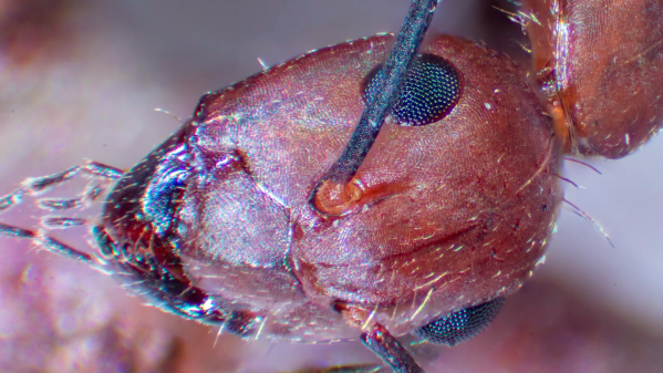
The code is very simple, and the setup is quite temporary looking, but the results are pretty impressive. We could do without the extreme closeup of that tick — nasty little arachnids — but the ant at the end of the video below has some interesting details. [ItMightBeWorse] doesn’t mention how the actual stacking is being done, but this CNC-based focus stacking project mentions a few utilities that take help with the post-processing.

Continue reading “Quick And Dirty Microscope Motion Control For Focus Stacking”

Your Phone Is A 200X Microscope — Sort Of

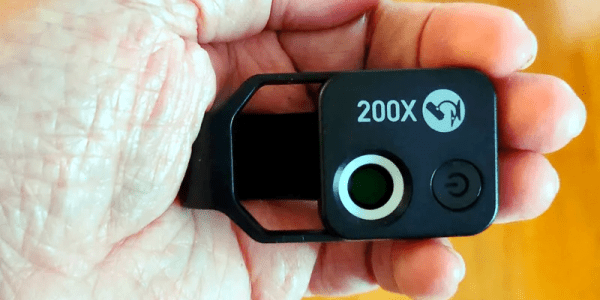
[A. Cemal Ekin] over on PetaPixel reviewed the Apexel 200X LED Microscope Lens. The relatively inexpensive accessory promises to transform your cell phone camera into a microscope. Of course, lenses that strap over your phone’s camera lens aren’t exactly a new idea, but this one looks a little more substantial than the usual piece of plastic in a spring-loaded clip. Does it work? You should read [Cemal’s] post for the details, but the answer — as you might have expected — is yes and no.

On the yes side, you can get some pretty neat photomicrographs from the adapter. On the negative side, your phone isn’t made to accommodate microscope samples. It also isn’t made to stay stable at 200X.

Continue reading “Your Phone Is A 200X Microscope — Sort Of”

Microscopy Hack Chat With Zachary Tong

Join us on Wednesday, June 23 at noon Pacific for the Microscopy Hack Chat with Zachary Tong!

There was a time when electronics was very much a hobby that existed in the macroscopic world. Vacuum tubes, wire-wound resistors, and big capacitors were all mounted on terminal strips and mounted in a heavy chassis or enclosure, and interfacing with everything from components to tools was more an exercise in gross motor skills than fine. Even as we started to shrink components down to silicon chips, the packages we put them in were still large enough to handle and see easily. It’s only comparatively recently that everything has started to push the ludicrous end of the scale, with components and processes suitable only for microscopic manipulation, but that’s pretty much where we are now, and things are only likely to get smaller as time goes on.

The microscopic world is a fascinating one, and the tools and techniques to explore it are often complex. That doesn’t mean microscopy is out of the wheelhouse of the average hacker, though. Zachary Tong, proprietor of the delightfully eclectic Breaking Taps channel on YouTube, has been working in the microscopic realm a lot lately. We’ve featured his laser scanning confocal microscope recently, as well as his latest foray into atomic force microscopy. In the past he has also made DIY acrylic lenses, and he has even tried his hand at micromachining glass with lasers.

Zach is pretty comfortable working in and around the microscopic realm, and he’ll stop by the Hack Chat to share what he’s been up to down there. We’ll talk about all the cool stuff going on in Zach’s lab, and see what else he has in store for us.

join-hack-chatOur Hack Chats are live community events in the Hackaday.io Hack Chat group messaging. This week we’ll be sitting down on Wednesday, June 23 at 12:00 PM Pacific time. If time zones have you tied up, we have a handy time zone converter.
Click that speech bubble to the right, and you’ll be taken directly to the Hack Chat group on Hackaday.io. You don’t have to wait until Wednesday; join whenever you want and you can see what the community is talking about.

Optical Microscope Resolves Down To 40 Nanometers

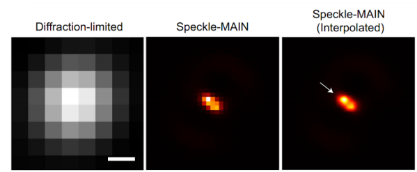
Optical microscopes depend on light, of course, but they are also limited by that same light. Typically, anything under 200 nanometers just blurs together because of the wavelength of the light being used to observe it. However, engineers at the University of California San Diego have published their results using a hyperbolic metamaterial composed of silver and silica to drive optical microscopy down to below 40 nanometers. You can find the original paper online, also.

The technique also requires image processing. Light passing through the metamaterial breaks into speckles that produce low-resolution images that can combine to form high-resolution images. This so-called structured illumination technique isn’t exactly new, but previous techniques allowed about 100-nanometer resolution, much less than what the researchers were able to find using this material.

Continue reading “Optical Microscope Resolves Down To 40 Nanometers”

High Tech Photos Capture Snowflakes Like Never Before

Microscopy used to be a rarity in the hobby electronics world. But anyone doing lab work has always needed a microscope and with today’s tiny parts, it is almost a necessity. However, [Nathan Myhrvold] didn’t use an ordinary microscope to capture some beautiful snowflake pictures. According to [My Modern Met], the pictures are the highest resolution snowflake pictures ever taken.

Of course, the site is more interested in the visual aspect of it, but they did provide some clues about the tech behind the pictures. According to the site:

Myhrvold used a special camera of his own design. He combined the magnifying power of a microscopic lens.. with a specially designed optical path. This path allowed the lens to channel its image to a medium-format digital sensor… In addition, the camera featured a cooling stage upon which the tiny specimens could rest. With LED short-pulse lights and a shutter speed of less than 500 microseconds, Myhrvold was able to capture multiple images of each snowflake at different focal lengths. These images were then stacked to create the final image.

Continue reading “High Tech Photos Capture Snowflakes Like Never Before”

Video: Bil Herds Looks At Mitosis

I loved my science courses when I was in Junior High School; we leaned to make batteries, how molecules combine to form the world we see around us, and basically I got a picture of where we stood in the  scheme of things, though Quarks had yet to be discovered at the time.

In talking with my son I found out that there wasn’t much budget for Science learning materials in his school system like we had back in my day, he had done very little practical hands-on experiments that I remember so fondly. One of those experiments was to look and draw the stages of mitosis as seen under a Microscope. This was amazing to me back in the day, and cemented the wonder of seeing cell division into my memory to this day, much like when I saw the shadow of one of Jupiter’s moons with my own eyes!

Now I have to stop and tell you that I am not normal, or at least was not considered to be a typical young’un growing up near a river in rural Indiana in the 60’s. I had my own microscope; it quite simply was my pride and joy. I had gotten it while I was in the first or second grade as a present and I loved the thing. It was just horrible to use in its later years as lens displaced, the focus rack became looser if that was possible, and dirt accumulated on the internal lens; and yet I loved it and still have it to this day! As I write this, I realize that it’s the oldest thing that I own. (that and the book that came with it).

Today we have better tools and they’re pretty easy to come by. I want to encourage you to do some science with them. (Don’t just look at your solder joints!) Check out the video about seeing mitosis of onion cells through the microscope, then join me below for more on the topic!

Continue reading “Video: Bil Herds Looks At Mitosis”

The Smallest Cell Phone Picture

Mobile phones are the photography tool for most of us, but they are a blunt tool. If you love astrophotography, you buy a DSLR and a lens adapter. Infrared photography needs camera surgery or a special unit. If you want to look closer to home, you may have a microscope with a CCD. Your pocket computer is not manufactured for microscopy, but that does not mean it cannot be convinced. Most of us have held our lens up to the eyepiece of some binoculars or a microscope, and it sort of works, but it is far from perfect. [Benedict Diederich] and a team are proving that we can get darn beautiful images with a microscope, a phone holder, and some purpose-built software on an Android phone with their cellSTORM.

The trick to getting useful images is to compare a series of pictures and figure out which pixels matter and which ones are noisy. Imagine someone shows you grainy nighttime footage from an outdoor security camera. When you pause, it looks like hot garbage, and you can’t tell the difference between a patio chair and a shrubbery. As it plays, the noisy pixels bounce around, and you figure out you’re looking at a spruce bush, and that is roughly how the software parses out a crisp image. At the cost of frame rate, you get clarity, which is why you need a phone holder. Some of their tests took minutes, so astrophotography might not fare as well.

We love high-resolution pictures of tiny things and that isn’t going to change anytime soon.

Thank you [Dr. Nicolás De Francesco] for the tip.