An intrinsic property of paintings, that makes them both wonderful and very annoying, is the fact that they are physical objects. Sometimes they survive across the ages as amazing artifacts of their era, but they are also susceptible to being lost and even destroyed. Sometimes this destruction is deliberate, such as when a painting is painted over.
Artists reuse canvas all the time — painting over what was already there. Sometimes they might be coerced by a client into altering a painting, or removing entire elements from a scene. Fortunately, nowadays we have many techniques, involving x-rays and infrared radiation, that can analyze paintings to determine not only the composition of what we can see with the naked eye, but also that what lies underneath.
In some cases, we can then reconstruct what was previously hidden, returning to physical reality paintings and sketches which haven’t seen the light of day for sometimes centuries.
Paintings are like onions
Painting is an additive method, which means that, starting with a blank canvas or other surface, paint is applied in one or more layers, which may overlap with other layers. When painting over an existing painting, the new paint tends to be applied on top of these existing layers. While this may obscure what was originally there from the human eye, it is possible to non-destructively peel back these layers, and analyze them individually.
The main methods to do this use x-rays and infrared. For decades, this meant using either creating an x-ray image from the entire painting, or illuminating the work with infrared light and capturing the reflected image. While this provides some level of detail that the naked eye cannot see, these methods cannot distinguish between layers of paint, nor details such as specific types of lead-based paints. Newer, more sophisticated methods can provide this kind of information.
A good overview of these methods comes from a paper by Stijn Legrand et al. (2014), as published in Heritage Science. Using the example of a 15th century triptych (folding three panel altar painting), it is demonstrated how this particular work saw multiple revisions over the years. These alterations primarily involved adding more of the family’s children, and moving existing figures around the painting to make space, which covered up the already painted-in background. Some of the female characters’ dresses were made more modest later as well, presumably after one of the daughters became a nun.
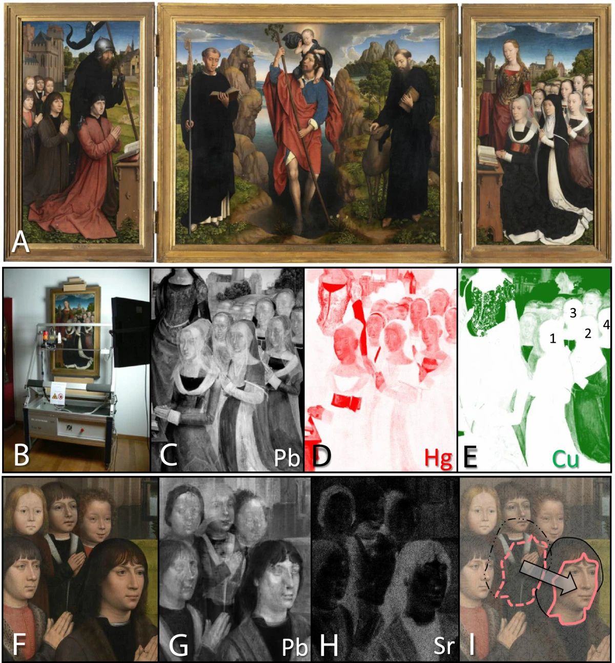
This information was gleaned from a macroscopic X-ray fluorescence (MA-XRF) scan, which uses X-rays to ionize the material. The radiation that is emitted from the material after being thus ionized is indicative of which elementary elements, like lead, are present, and in which quantities. As each type and color of paint is made up out of different mixtures of these elements, this allows us to see hidden layers as a kind of ghost image despite MA-XRF not being layer sensitive.
While exceedingly useful, there is a way to also get the exact composition of the paints used: macroscopic X-ray diffraction (MA-XDR). Instead of tracking the fluorescence using a sensor placed behind the painting, this method puts a sensor at an angle with the front, alongside the X-ray source:
![Prototype MA-XRD setup at the University of Antwerp. A) Photograph showing the micro-focus X-ray tube source (S), equipped with a double curved mirror M and detector for recording transmission XRD (D1) and XRF (D2) data: these components are positioned close to a painting mounted on a motorized stage; B) MA-XRD and C) MA-XRF images obtained by scanning a detail of the painting shown in D): scan size: 78×75 mm2, image step size: 0.5 mm in both directions, Dwell time: 2 s/pixel. Adapted from [Vanmeert F, Janssens K, De Nolf W, Legrand S, Van der Snickt G, Dik J: Scanning Macroscopic X-ray powder diffraction imaging (MA-XRPD): transfer from the synchrotron to the laboratory, submitted]. (Source: Stijn Legrand et al., 2014)](https://hackaday.com/wp-content/uploads/2021/12/40494_2014_Article_40_Fig3_HTML.jpg)
Peeling Back Layers
But let’s go deeper. What all of the previously mentioned X-ray and IR-based methods have in common is that they do not distinguish between the individual layers of the painting. But just as you can take X-rays of people from multiple angles, you can also use Computed Tomography (CT) scans on art.
For a traditional CT scan it is of course essential that either the scanner or object rotates so that radiographs can be made of as many angles as possible. While this works well for basically round human bodies and similar shapes, flat objects like paintings and panels of composite materials cannot be scanned in this manner. Here we see that over the past years there’s been a flurry of research from both the art and materials research fields into ways to adapt traditional CT scans into a form that will work with these flat objects. This gets us to Computed Laminography (CL).
Charles E. Wood et al. (2019, PDF) report on two different ways to perform CL on carbon fiber reinforced polymer (CFRP) sections. The goal here is to detect defects in the composite material, such as air bubbles and fractures. Using both a hexapod and robotic arm setup, CL scans were made, following both a raster and a limited angle scan pattern (as opposed to the full rotation of CT). The data was then assembled into the computed model using the Simultaneous Iterative Reconstruction Technique (SIRT) iterative reconstruction algorithm.
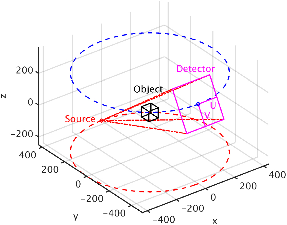
object’s frame of reference xyz. (Source: S.L.Fisher et al., 2019)
While this gave usable results, the imprecise positioning and reduced angles introduced artifacts in the final model. S. L. Fisher et al. (2019) reference Wood et al. in a study on an approach using CT scanning equipment as found in laboratories and using a single rotation axis for the subject.
This approach seems to get around many of the limitations of both CT and many of the CL configurations. By allowing for full rotation of the object being scanned, this allows for essentially the full range of angles, while simultaneously offering a stable platform without the positional difficulties encountered by Wood et al.
Although still just a proof-of-concept study, the approach shown by Fisher et al. shows that there are fairly straightforward ways to implement CL, without the use of expensive equipment or a complicated scanning setup.
Restoring That Which was Lost
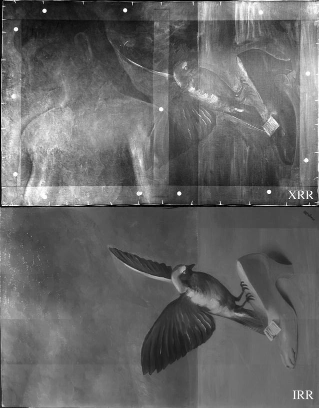
Over the years, methods like these have been used by art conservators and others to not only understand the physical make-up of a painting, but also the techniques used. With CL in particular, it becomes possible to see the brush strokes and layering used to achieve particular effects. By using XRD to analyze the specific pigment used in an overpainted section, it is possible to even reconstruct the exact color of that section. This was essential with the reconstruction of La pose enchantée by René Magritte (1898–1967), as documented by Catherine Defeyt et al. (2018)
Created in 1927, this painting vanished around 1932. It was known that Magritte’s financial situation led him to often reuse old works. By examining other works by this artist, it was found during X-ray and IR-based scans that this vanished painting had in fact been cut up and used for new works.
Reconstructing this work involved scanning existing paintings to find the individual pieces, matching this up with a monochrome photo of the work taken before it was cut up and using XRF to determine the pigments used for the original painting layers. This information was then combined to reconstruct what the original, full-color painting would have looked like. Even though this will not bring back the original work, it does give us an in-depth look at its original appearance.
Keeping It Real
In the world of art, everything is about authenticity. This becomes quite evident every time a ‘previously unknown’ or ‘assumed lost’ work of some master surfaces and the debate about whether it’s really by this painter begins. An interesting aspect of this is whether a reconstructed, physical version of a previously lost copy has anything in common with the original. With recent reports of for example a lost Picasso being reconstructed using a neural network trained on other works by Picasso, it’s perhaps best to see these as reproductions.
A few years ago the Smithsonian Magazine covered the reconstruction of seven lost masterpieces. This effort used any existing photographic and eyewitness evidence along with knowledge about the painter’s techniques to paint these works anew. Even though we know that these works are as close to the original as possible, we also know that they were not truly painted by Van Gogh, Monet or any of the other masters.
Even so, there is a lot to be said for having a physical copy of a piece of history that was once assumed to be lost forever, no matter for how long the debate about where the true value of an art piece lies.


Thank you for the interesting work being done in the study off old art.
I also remember how the progression of written historical documents is being lost through the use of word processors. In today’s documents, their is no “paper trail” of edits done during the creation of the document. I guess the same could be said for pictures whole developed using software, unless incremental backups are preserved.
Well in the case of Hackaday comments, edits aren’t allowed, so our great works of writing are preserved. B^)
Blockchain ain’t got nothing nothing on us!
PS: Noticed reply contained error. Left it as-is out of solidarity with y’all.
Thanks thanks.
I think this is a feature. If one _really_ scews up a comment, one can make another, to correct the first. Knowing there is no edit feature makes me pause before submitting, which (hopefully) encourages proofreading and more thoughtful posts.
Actually, many formats like office documents do keep an edit trail, similar to a journal file. This is however periodically cleared, or saving to a new file also clears it.
We’ve got git.
Well even with git there are ways to avoid showing full history.
This was my first thought also.
Despite being a purely scientific endeavor, this is magic. X-rays and math, sure… but it’s magic. It confounds me that such feats are possible and yet I understand the process.
What concerns me is that those paintings contain mercury, lead and other toxic materials. They should treated as hazardous waste and recycled. If their content is still relevant it could be replaced by digital reproductions.
The elements have to exist somewhere. What’s wrong with having them contained within a work of art? It’s not like little children are chipping off the paint and eating it. What would recycling even mean? Like, turn the painting into a NiCad battery? Bury it in a landfill?
Thirty-odd years ago, I stayed in a hotel that had a “haunted” painting. There was a shadowy figure that could be made out alongside the other figures in the painting, which the hotel staff said had not been present in the painting when it was first hung. I always presumed that it was one that the artist had originally put in, but decided to paint over for whatever reason.
It would be interesting to have these types of scan done on that painting, but I suspect the hotel would prefer to have a “haunted” painting.
Perhaps the missing person is Trotsky?
B^)
“In the world of art, everything is about authenticity.” Nope, that would be the world of investment in objects, artefacts. In the world of Art everything is about original ideas and perceptions or experiences. Using a computer to take the style of a living artist and apply it to a photographic portrait of them is also Art, if you are making a point about what it implies to do such a thing. I did it to Damien Hirst once, the poor guy probably started to question his intake of inebriants that day. The point or the Art of doing such a thing was a commentary on the fight Hirst got into with indigenous Australians because of his series of dot paintings.
Ceci n’est pas une 555.
If I’d been drinking coffee I would need a new keyboard now.
You win Hackaday.
Hmm… we’re hitting one of a kind works of art with IONIZING radiation? I’m all for using clever math and waves to learn stuff, but wonder how close this gets to changing the thing we’re looking at.
Not very close at all.
Just don’t try it with comic book art. Never expose comic book art to radiation.
Because that is how we got Spiderman and Hulk!
How I’d do CT of a flat item like a painting would be to lay the painting flat on a turntable then angle the X-Ray projector at it, with the detector behind. Spin the turntable and run the X-Ray projector inward in steps like tracks on a floppy disk. Go back to the start, slightly change the beam angle then go again. Make 100 or so passes with the beam angle different every pass.
A very nice interactive website was created (in cooperation with University of Antwerp and Brussels) about the Altarpiece of Van Eyck where the X-rays (and more, like infrared) are also available:
http://closertovaneyck.kikirpa.be/