[Pyrotechnical] thought about buying a CAT scanner and found out they cost millions of dollars. So he decided to build one for about $200 using a salvage X-ray tube and some other miscellaneous parts. A scintillating detector provides the image for pick up with a camera phone. The control? An Arduino, what else? You can watch the video below, but due to plenty of NSFW language, you might want to put your headphones on if you don’t want to shock anyone.
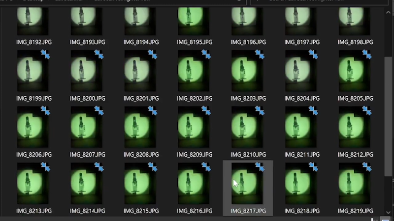
Of course, you need to be careful when working with energetic X-rays. To keep away from the X-ray source, [Pyrotechnical] used a Roku remote and an IR sensor to control the device from afar. The electronics is pretty easy. You just have to rotate a turntable and trigger the camera while lighting up the X-ray tube.
The real problem is performing tomography (the T in CAT) to convert the flat pictures into a 3D representation. There’s software for that, of course, and that’s what he uses. Honestly, this reminded us of the cheap 3D scanners that use a cell phone and a turntable. If you took one of those and added the scintillator and the X-ray tube, it should work just as well.
If you decide to replicate this, please be sure you understand X-ray safety. You shouldn’t be subjecting anything alive to the beam — cat or otherwise. The “throw-together” build quality reminded us of another homemade X-ray machine we’ve seen. Surprisingly, this isn’t the first homemade CAT scanner we’ve seen.
(Reminder, the video below contains a good bit of NSFW language.)


Without a Radon transform this is hardly a CT scanner. Sorry for being so blunt, but I got hyped first, only to be disappointed to just see classical xrays being performed. No sinograms spotted!
Use open of the projects on github ?
https://github.com/topics/radon-transform
“Use one …”, brain is telling me it is coffee time.
Here is another approach to turn CT into actual 3D. Still need to create enough data to begin with though.
https://www.youtube.com/watch?v=WBk9Z-CgJeg
[youtube https://www.youtube.com/watch?v=WBk9Z-CgJeg&w=560&h=315%5D
I mean there’s a 3d point cloud at the end and he’s able to slice the object into layers. That’s computed tomography.
Did you watch the video around 10 minutes. He uses a program to do cone-beam backprojection from the X-rays. I’m no expert, but I think it qualifies as CT.
My diagnostic radiology class was long, long ago so I may be incorrect. But. Tomography is the process of getting a flat image out of a 3d structure, not the other way around. As in, a “virtual” slice through, say, a head. It is very difficult to explain, but before the computed tomography scanners, you could still do tomography with just a beam and a flat x-ray plate. I’m sure a quick google search will get you an image that is worth my 1000 poorly chosen and confusing words. What some are talking about below is what would be termed “3d reconstruction” and is not really tomography. That is a very complex process of, for example, injecting contrast media, spinning the CT scanner rapidly around the brain to take many x-rays that are then processed (computed) into flat plate images, that are then processed again into a virtual stack, the other stuff filtered out, leaving you with a neat movie of the contrast enhanced cerebral vasculature. Solving the tomography algorithm using FFT was a major medical imaging breakthrough. Again, all specific examples of the general process.
I’m pretty sure at the end he shows the slices of the 3d construction. The very end
This is the approach known as cone beam computed tomography is used extensively in the medical industry. Particularly where there are size and weight constraints on the equipment for modalities where x-ray CT is a secondary function and been essentially bolted on to the gantry’s i.e. Single Photon computed tomography (SPECT),This CT data is then fed into the SPECT system for attenuation corrections to improve image quality. This is usually done for mobile (as in the back of a HGV) or for on radiotherapy treatment linear accelerators for image matching, it has the added benefit of being able to take planar images in addition to CBCT. The image quality is sub par and imaging doses to the patients are significantly higher compared to a CT scanner using a fan beam as scattered photons from within tissue creates noise across the imaging panel (requiring a much higher dose, i.e. beam current to get a comparable single to noise) the longer imaging time (there is a IEC spec for none enclosed gantry’s having a limit of 1RPM) this leads to other imaging artefacts as in temporal blurring due to patient motion i.e. respiration, where a fan beam CT scanner is usually 2RPS, 120RPM.
TLDR: This is a CT technique, just not typically used diagnostically.
Has anyone here tried (successfully) put together an medical ultrasound rig (capable of producing usable images) ? If yes please share.
I’ve seen cheap ultrasound imagers sold for pregnant women to do their own imaging for a few hundred dollars but have never looked into it
They have done this for breast cancer detection using video cards in a desktop system for many years, but they still put your chesticles in the big squishy device. The big squishy device takes around two or four weeks to get results, last i heard the sonogram version using video cards for imaging took fourty five minutes. Very old data i read about countless years ago, back when sli was still a thing.
These exist?
We only used one of those cheap Cardiotocography machines from China. I bought it cheap on Aliexpress and after my wife gave birth we sold it locally, at small profit. It displayed the heart rate and played the heart beat. IIRC, it used Doppler shift created by moving heart to create beat frequency from the ultrasound generated at few MHz. Also it works with adult hearts and can play sounds of digestion.
That’s what we usually just call the Doppler. It is a probe that uses doppler ultrasound to measure blood flow (typically).
To answer the original poster’s question, I haven’t seen anyone try to make their own ultrasound, but the but the cost of commercial ones is coming way down. Ones used in hospitals for echocardiography can be $100k’s, more typical models a bit less at 40-50k. For “only” $2k you can get a svelte one that plugs into your iPhone or Android. I’ve tried some other manufacturers’ offerings but that one is the best.
I just found an used ultrasonograph, Toshiba Xario SSA-660A with added optional upgrades for equivalent of ~350USD. Refurbished Toshiba Aplio TUS-A300 with upgraded firmware and few different heads goes for about ~17150USD. The most expensive new one I found was Acclarix AX3 for about 10000USD. However I didn’t check medical suppliers on their sites. Still I’m under the impression that anything medical in USA is much more expensive than in Poland, or other European countries.
@Arthur said: “Has anyone here tried (successfully) put together an medical ultrasound rig (capable of producing usable images)? If yes please share.”
A 1D Doppler ultrasound might be easy to make, but I have never seen one. I have seen a 3D ultrasound wand that plugs into a smart phone or tablet though. The Mobisante MobiUS SP1 Smartphone Ultrasound wand costs £7,000 ($8,452.50) compared to £60,000 ($72,450.00) for a full-size ultrasound.[1] Unfortunately the Mobisante web site [2] doesn’t respond and from what I’ve seen they only have five employees.[3] There is a YouTube video of the device in action though.[4]
* References:
1. The ultrasound scanner that plugs into a SMARTPHONE and could revolutionise medical care in third world countries
https://www.dailymail.co.uk/sciencetech/article-2363964/The-ultrasound-scanner-plugs-SMARTPHONE-revolutionise-medical-care-world-countries.html
2. Mobisante web site (Kaput)
https://www.mobisante.com/
3. Mobisante Company Info
https://gust.com/companies/mobisante
4. Ultrasound machine that fits in your pocket: The smartphone-based MobiUS [YouTube 00:54]
https://www.youtube.com/watch?v=MX8jTUqnaAs
You can occasionally find old Merlin/Ecklin cards and transducers for sale cheap or free, in need of repairs or software. That’s bottom of the barrel tech but with a little work you can measure a kidney or identify a change in tissue density or identify a fetus. Self-contained 4d units start around $2500 but you get dedicated physical controls (which is handy.)
If you want to scan medical implant castings for defects, my brother-in-law does that on a $5m+ rig. He estimates he rejects more than half of the hips and knees produced due to voids or irregular crystals in the titanium.
5 millidollars?
B^)
Hot take: life saving devices should never be subject to millions of dollars in licensing fees.
This is a good case for funding nasa rather than spacex as the things nasa engineerds invent become public domain and the things spacex invents will be patented for all time if yhey have their way. Just one example. Id rather pay tax to fund nasa than pay tax to fund any private entity. Period.
Would you rather pay 1 billion dollars for NASA to get something from their ‘usual channels’ or 50 million dollars for NASA to get something from SpaceX?
The ‘usual channels’ all become jobs programs. The work has to be split across all congressional districts and the patients still end up in private hands. The money just circles back and makes government even more corrupt (barely possible).
I don’t think you know how government contracting works. They didn’t want to give SpaceX money. Many more BJs and drinks available from the usual suspects.
Trust me on this, I’ve taken many public utility managers and their wives out for really expensive dinners. Nudie bars and worse too, just less the wives. Many nations, not just USA.
Free lifesaving devices isn’t one of your choices. It’s pay for it or not have it. Alternatively you can pay for it and still not have it, but congressional nephews get gravy.
@dudenamedben said: “Hot take: life saving devices should never be subject to millions of dollars in licensing fees.”
Life saving devices are usually complex, therefore costly to develop and build. Also the insurance premiums the medical equipment manufacturers must pay to defend themselves against persistent attacks from greedy Trial Lawyers are astronomical. Finally, money doesn’t grow on trees! So the money has to come from somewhere, and licensing fees are one source. But ultimately the money always ends up coming from the end-users and taxpayers.
How about a massive tax on junk food to subsidize healthcare?
The main barrier is not the license tied to life-saving devices, but the liability tied to them. A Xray machine can malfunction and give someone cancer, or don’t malfunction and someone gets cancer anyway. User sues, how will pay the bill?
The hefty price is the amount of certifications, safety measures, safety studies, insurance and everything related to it. You can create an open source CT machine with parts bought on eBay and no hospital is going to use it anyway. Because when it malfunctions somehow, you aren’t there to pay reparations to any injured person.
For the hospital, a free CT machine without a solid company behind can end up way more expensive than a half million one from Siemens. A single malfunction and every patient that used the machine during certain timeframe will have to be contacted, monitored, and if they have any health issues they will have to be compensated. Have this scenario with 20 patients all asking for 6-digits reparations and that 500k Siemens CT is a hell of a bargain.
Seems like Tesla also experimented with x-rays / shadowgraphs. Back in the 1800’s. Was probably a bit more complicated back then. Let alone as distantly treated or shielded as they are today!
https://teslauniverse.com/nikola-tesla/images/x-ray-photograph-called-shadowgraph-tesla-teslas-foot
Quote: ‘you need to be careful when working with energetic X-rays.’
You need to be doubly careful when working with soft x-rays. As used in x-ray crystallography. Much more of the energy is absorbed. The beam is death.
True, the lower energy x-ray photons used in crystallography do not penetrate as deeply into body tissue, but a blanket statement like that is pretty misleading.
Would you rather have your body absorb 100% of the 20 keV photons from a 50 watt crystallography source, or 90% of the 80 keV photons from a 60 kilowatt diagnostic x-ray tube? The diagnostic tube deposits 4000 times more radiation dose into your body.
Now, if you stick your finger on the output of the crystallography tube for a significant length of time, you’ll probably regret that life choice, but it wouldn’t be ‘death’.
My dad did crystallography.
Your absorption ratios and power levels are way off.
If you intercept the beam of a crystallography machine with a body part the best option is amputation. Gangrene is coming quickly.
Do you have numbers?
In the first centimeter of human tissue, the beam from a crystallography Cu source (9 keV) deposits about twice as much energy (or radiation dose) per unit of beam power compared to a chest x-ray or CT scanner’s 80 keV energy. But the lower energy photons stop there. 80 keV photons barrel on for several more centimeters, but even those are mostly stopped and deposit their radiation dose in human-size objects. Only 3% make it through an abdomen, for example.
So even though a crystallography source doses the first few millimeters of tissue at a rate twice as high as diagnostic x-ray energies, an 80 keV photon deposits 8x more dose in a body.
A microfocus crystallography tube is 50-80 watts, or up to a couple of kilowatts for the large-focus devices. A CT or chest x-ray tube typically runs around 60 kilowatts, 30-1000 times more power.
Now, in diagnostic imaging you are 0.7 m (CT) up to 2 meters (chest x-ray) from the source, and exposure times are just a few seconds (CT) or a tenth of second (chest). Despite the distance, dose rates are high. An hour of continuous exposure would be enough to kill you (it would melt the tube long before that, though).
But the inverse-square law works both ways: Even if the total energy from a crystallography source is low, If you’re unlucky (or dumb) enough to linger close to it for an extended period, you’re absolutely right: that body part can easily receive a lethal dose.
It’s not so much the photon energy that does it as the distance from the source. Soft x-ray photons deposit only a few times more energy than their high-energy cousins, but it’s easy to get very close to the source: enough so that even a low power source can be very hazardous.
Don’t remember the details, dad was the physicist. Just lowly BSEE (to dad’s great disappointment). Grew up on campus, wasn’t going to get near that mess. I was down the road at the kegger.
IIRC it was the focused nature of the soft x-ray beam (combined with it’s ready absorption in a shallow area). No inverse squared law in play. Didn’t affect much volume, but nuked what it did.
Recall a series of pictures of an incident he had. Started out looking like a dark freckle on the guys hand. Ended with arm stub.
Also something P-chemish about breaking bonds at soft x-ray energy.
This was all decades ago. Machines are likely much safer now.
This one was like my 1950 sickle bar mower (aka ankle mower). Better not be stupid.
Dad was also the guy who taught me how to make LOX out of liquid N (liquid air intermediate step). Years later I saw the burn mark on the bar table at Heidelberg where he had soaked a rope in LOX, lit it and swung it around his head. Then decided it was wise to let it go. I know where my explosive pyromania comes from.
I digress. Miss him. Think I’ll blow something up in his memory.
You weren’t kidding about the gratuitous NSFW language. I wonder if it’s an after-effect of his previous head injury. It sure doesn’t make him sound like a grown-up, if that was the effect he was aiming for…
+1 for ingenuity, -1 for language.
10 minute exposures with no filtration/beam hardening, no shielding other than distance and no visible dosimeters… Nothing stopping the primary beam but the image receptor. I hope nobody else is home or closer to that tube than he is. I really hope nobody tries this in an apartment.
Unless he is working in an environment that is properly shielded, he is retarded and a danger to his community.