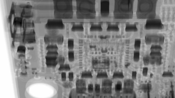
Fluoroscopy is probably the best-known method of X-ray imaging: an X-ray beam passes through the subject to be imaged, and the transmitted X-rays illuminate a phosphor screen. Dense objects, such as metal or bone, cast a shadow on the screen, which provides a real-time image of the subject’s interior. Already having access to X-ray sources, [MarcellF]’s next step was to investigate common phosphor materials, then synthesize his own.
Most common materials that fluoresce under ultraviolet light showed no activity under X-rays: fluorescein, quinine, UV fluorescent paint, and common fluorescent minerals emitted no noticeable glow under 80 kV X-ray stimulation. However, strontium aluminate phosphors did fluoresce well, with a strong afterglow, as did the phosphors in a fluorescent light bulb, some LEDs, and an electroluminescent panel. The electroluminescent panel, which used a zinc sulfide phosphor, was almost as bright as the gadolinium oxysulfide screen from a CT scanner’s detector and had no noticeable afterglow.