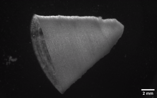
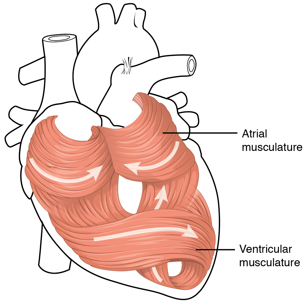
What makes a body’s organs into what they are is more than just a grouping of specialized cells. They also need to be oriented and attached to each other and scaffolding in order to create structures which can effectively perform the desired function. A good example here is the heart, which requires a large number of muscle cells to contract in unison in order for the heart component (like a ventricle) to effectively pump blood. This complication is what has so far complicated efforts to 3D print complex tissues and entire organs, but recently researchers have demonstrated a way to 3D print heart muscle which can contract when stimulated similarly to a human heart’s ventricle.
At the center of this technique lies a hydrogel that is infused with gelatin fibers. Using a previously developed Rotary Jet-Spinning technology that was reported on in 2016, a sheet of spun fibers was produced that were then cut up into micrometer-sized fibers which were dispersed into the hydrogel. After printing the desired structure – taking into account the fiber alignment – it was found that the cardiomyocytes (the cells responsible for carrying the contractile signal in the heart muscle) align along the thus laid out pattern, ultimately creating a cardiac muscle capable of organized contraction.
These findings come after many years of research into the topic, with e.g. Zihan Wang and colleagues in a 2021 paper reporting on the challenges remaining with 3D printing cardiac tissue, yet also the massive opportunities that this could provide. Although entire heart replacements (via therapeutic cloning with the patient’s own cells) might become possible too, more immediate applications would involve replacements for damaged cardiac muscle and other large structures of the heart.


As a Hypertrophic cardiomyopathy patient, this is exciting news.
Welp, gotta support it until it turns 18.
Abortion legal to 87th trimester or 4 trimesters after the last time they ask their parents for money!
They are just a helpless cluster of cells at that age.
I just did a lit review and then thought of the new likely to work first ever blood alternative under clinical trials and the costing the surface with stem cells and let it adhere before implanting. But they are already using and testing diffrent formulations. One mixture contains hydrogel.
I wrote the corresponding author about my idea as using this on facial part could speed, of the idea works,.it’s use in extreme cases where death will likely occur otherwise and I am a medical researcher;
Here is the full current abstract form 2023:
Satar Yousefiasl, Esmaeel Sharifi, Erfan Salahinejad, Pooyan Makvandi, Soussan Irani,
Bioactive 3D-printed chitosan-based scaffolds for personalized craniofacial bone tissue engineering,
Engineered Regeneration,
Volume 4, Issue 1,
2023,
Pages 1-11,
ISSN 2666-1381,
https://doi.org/10.1016/j.engreg.2022.09.005.
(https://www.sciencedirect.com/science/article/pii/S2666138122000615)
Abstract: Regeneration of craniofacial bone defects is a key issue in the bone regeneration field. Hence, novel treatment strategies, such as tissue engineering using porous scaffolds, have been developed. An ideal tissue-engineered scaffold for bone tissue regeneration should possess pores to facilitate nutrients transmission and support reparative tissue ingrowth, bioactivity for osteoconduction and osseointegration, and biocompatibility to improve cell attachment, proliferation, and extracellular matrix formation. In the present study, we manufactured chitosan-based hydrogels substituted with alginate with optimized properties by extrusion-based three-dimensional (3D) printing. 3D printing of the scaffolds enables the designing and developing of complex architectures for craniofacial reconstruction using computer-aided design (CAD). Different ratios (2.5, 5, and 10%) of hydroxyapatite were added to the hydrogel, printed, and subsequently lyophilized to augment the physical and biological characteristics of the scaffolds. Hydroxyapatite incorporation into the chitosan-based scaffolds increased the porosity and pore size of the printed scaffolds. In addition, the presence of hydroxyapatite amplified apatite formation and decreased the size of formed apatite crystals. All the scaffold samples showed biocompatible properties and did not have toxicity toward rat bone marrow mesenchymal stem cells. Furthermore, the scaffolds containing 5% w/w hydroxyapatite exhibited significant growth in cell viability compared to the control. Overall, it is concluded that chitosan-based scaffolds adorned with hydroxyapatite are considerable for regenerating craniofacial bone defects.
Keywords: Bioactivity; CAD/CAM; Maxillofacial reconstruction; Pore; Stem cells
I did not read all of this, but, this appears related to similar research I had done many years ago with 3d printed silicon covered with a “human paste” stem cells like chia pet seeds and watched skin cells grow.
First thought… That’s terrible. I can even think of adults that would still fit that description.
Second thought.. I can think of some adults that still fit that description. Great idea!!
Face Off for real comes to mind! Nicolas Cage and John Travolta – ? Yes?