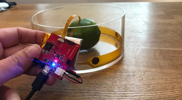
We live in a world where anyone can build a CT machine. Yes, anyone. It’s made of laser-cut plywood and it looks like a Stargate. Anyone can build an MRI machine. Of course, these machines aren’t really good enough for medical diagnosis, or good enough to image anything that’s alive for that matter. This project for the Hackaday Prize is something else, though. It’s biomedical imaging put into a package that is just good enough to image your lungs while they’re still in your body.
The idea behind Spectra is to attach two electrodes to the body (a chest cavity, your gut, or a simulator that’s basically a towel wrapped around the inside of a beaker). One of these electrodes emits an AC signal, and the second electrode measures the impedance and phase. Next, move the electrodes and measure again. Do this a few times, and you’ll be able to perform a tomographic reconstruction of the inside of a chest cavity (or beaker simulator).
Hardware-wise, Spectra uses more than two electrodes, thirty-two on the biggest version built so far. All of these electrodes are hooked up to a PCB that’s just under 2″ square, and everything is measured with 16-bit resolution at a 160 kSPS sample rate. To image something, each electrode sends out an AC current. Different tissues have different resistances, and the path taken through the body will have different outputs. After doing this through many electrodes, you can use the usual tomographic techniques to ‘see’ inside the body.
This is a remarkably inexpensive way to image the interior of the human body. No, it doesn’t have the same resolution as an MRI, but then again you don’t need superconducting electromagnets either. We’re really excited to see where this project will go, and we’re looking forward to the inevitable project updates.


