3D Printing Livers

The University of Utrecht has a team that is successfully bioprinting “liver units” that are able to do some of the functions of a human liver and may open the door to new medical treatments. This isn’t simply printing a fake liver in a jar though, instead the technique uses optical tomography to rapidly create small structures of about 1 cc of volume in less than 20 seconds.

Apparently, one problem with printing hydrogels full of biological structures is that passing them through a nozzle tends to disturb the delicate structures.  This technique uses no nozzle or layers, which makes it useful in this situation.

Continue reading “3D Printing Livers”

Ask Hackaday: What Can Only A Computer Do?

It is easy to apply computers to improve things we already understand. For example, instead of a piano today, you might buy a synthesizer. It looks and works — sometimes — as a piano. But it can also do lots of other things like play horns, or accompany you with a rhythm track or record and playback your music. There’s plenty of examples of this: word processors instead of typewriters, MP3 players instead of tape decks, and PDF files instead of printed material. But what about something totally new? I was thinking of this while looking at Sonic Pi, a musical instrument you play by coding.

But back to the keyboard, the word processor, and the MP3 player. Those things aren’t so much revolutionary as they are evolutionary. Even something like digital photography isn’t all that revolutionary. Sure, most of us couldn’t do all the magic you can do in PhotoShop in a dark room, but some wizards could. Most of us couldn’t lay out a camera-ready brochure either, but people did it every day without the benefit of computers. So what are the things that we are using computers for that are totally new? What can you do with the help of a computer that you absolutely couldn’t without?

Continue reading “Ask Hackaday: What Can Only A Computer Do?”

Tomography Through An Infinite Grid Of Resistors

One of the vast untapped potentials of medicine is the access to imaging equipment. A billion people have difficulty getting access to an x-ray, and that says nothing about access to MRIs or CAT scans. Over the past few years, [Jean Rintoul] has been working on a low-cost way to image the inside of a human body using nothing more than a few electrodes. It can be done cheaply and easily, and it’s one of the most innovative ways of bringing medical imaging to the masses. Now, this is a crowdfunding project, aiming to provide safe, accessible medical imaging to everyone.

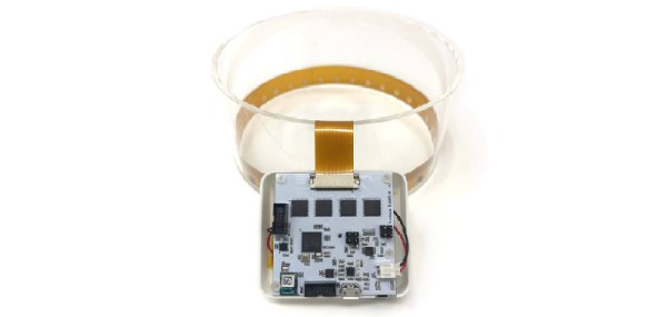
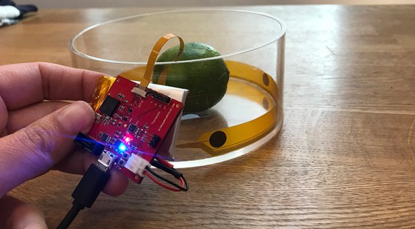
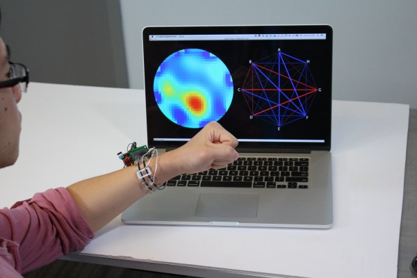
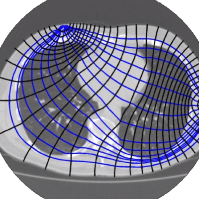
It’s called Spectra, and uses electrical impedance tomography to image the inside of a chest cavity, the dielectric spectrum of a bone, or the interior of a strawberry. Spectra does this by wrapping an electrode around a part of the body and sending out small AC currents. These small currents are reconstructed using tomographic techniques, imaging a cross-section of a body.

[Jean] gave a talk about Spectra at last year’s Hackaday Superconference, and if you want to look at the forefront of affordable medical technology, you needn’t look any further. Simply by sending an AC wave of around 10kHz through a body, software can reconstruct the internals. Everything from lung volume to muscle and fat mass to cancers can be detected with this equipment. You still need a tech or MD to interpret the data, but this is a great way to bring medical imaging technology to the people who need it.

Right now, the Spectra is up on Crowd Supply, with a board that can be configured to use 32 electrodes. Measurements are taken at 160,000 samples/sec, and these samples have 16-bit resolution. This is just the acquisition hardware, though, but the software to do tomographic reconstruction is open source and also readily available.

In terms of bringing medical imaging to the masses, this is a very impressive piece of work, and is probably the project from last year’s Hackaday Prize that has the best chance of changing the world.

3D Printing With Tomography In Reverse

The 3D printers we’re most familiar with use the fused deposition process, in which hot plastic is squirted out of a nozzle, to build up parts on a layer by layer basis. We’ve also seen stereolithography printers, such as the Form 2, which use a projector and a special resin to produce parts, again in a layer-by-layer method. However, a team from the University of North Carolina were inspired by CT scanners, and came up with a novel method for producing 3D printed parts.

The process, as outlined in the team’s paper.

The technique is known as Computed Axial Lithography. The team describe the system as working like a CT scan in reverse. The 3D model geometry is created, and then a series of 2D images are created by rotating the part about the vertical axis. These 2D images are then projected into a cylindrical container of photosensitive resin, which rotates during the process. Rather than building the part out of a series of layers in the Z-axis, instead the part is built from a series of axial slices as the cylinder rotates.

The parts produced have the benefit of a smooth surface finish and are remarkably transparent. The team printed a variety of test objects, including a replica of the famous Thinker sculpture, as well as a replica of a human jaw. Particularly interesting is the capability to make prints which enclose existing objects, demonstrated with a screwdriver handle enclosing the existing steel shank.

It’s a technique which could likely be reproduced by resourceful makers, assuming the correct resin isn’t too hard to come by. The resin market is hotting up, with Prusa announcing new products at a recent Makerfaire. We’re excited to see what comes next, particularly as the high cost of resin is reduced by economies of scale. Video after the break.

[via Nature, thanks to Philip for the tip!]

Continue reading “3D Printing With Tomography In Reverse”

Towards Open Biomedical Imaging

We live in a world where anyone can build a CT machine. Yes, anyone. It’s made of laser-cut plywood and it looks like a Stargate. Anyone can build an MRI machine. Of course, these machines aren’t really good enough for medical diagnosis, or good enough to image anything that’s alive for that matter. This project for the Hackaday Prize is something else, though. It’s biomedical imaging put into a package that is just good enough to image your lungs while they’re still in your body.

The idea behind Spectra is to attach two electrodes to the body (a chest cavity, your gut, or a simulator that’s basically a towel wrapped around the inside of a beaker). One of these electrodes emits an AC signal, and the second electrode measures the impedance and phase. Next, move the electrodes and measure again. Do this a few times, and you’ll be able to perform a tomographic reconstruction of the inside of a chest cavity (or beaker simulator).

Hardware-wise, Spectra uses more than two electrodes, thirty-two on the biggest version built so far. All of these electrodes are hooked up to a PCB that’s just under 2″ square, and everything is measured with 16-bit resolution at a 160 kSPS sample rate. To image something, each electrode sends out an AC current. Different tissues have different resistances, and the path taken through the body will have different outputs. After doing this through many electrodes, you can use the usual tomographic techniques to ‘see’ inside the body.

This is a remarkably inexpensive way to image the interior of the human body. No, it doesn’t have the same resolution as an MRI, but then again you don’t need superconducting electromagnets either. We’re really excited to see where this project will go, and we’re looking forward to the inevitable project updates.

Everything’s A Touch Surface With Electrick

Touch screens are great, but big touchscreens are expensive and irregular touchscreens are not easy to make at all. Electrik is a method developed by several researchers at Carnegie Mellon University that makes almost any solid object into a touch surface using tomography. The catch is that a conductive coating — in the form of conductive sheets, 3D plastic, or paint — is necessary. You can see a demonstration and many unique applications in the video below. They’ve even made a touch-sensitive brain out of Jell-O and a touchable snowman out of Play-Doh.

The concept is simple. Multiple electrodes surround the surface. The system injects a current using a pair of electrodes and then senses the output at the other terminals. A finger touch will change the output of several of the electrodes. Upon detection, the system will change the injection electrodes and repeat the sensing. By using multiple electrode pairs and tomography techniques, the system can determine the location of touch and even do rough motion tracking like a low-resolution touch pad mouse.

Continue reading “Everything’s A Touch Surface With Electrick”

Impedance Tomography Is The New X-Ray Machine

Seeing what’s going on inside a human body is pretty difficult. Unless you’re Superman and you have X-ray vision, you’ll need a large, expensive piece of medical equipment. And even then, X-rays are harmful part of the electromagnetic spectrum. Rather than using a large machine or questionable Kryptonian ionizing radiation vision, there’s another option now: electrical impedance tomography.

[Chris Harrison] and the rest of a research team at Carnegie Mellon University have come up with a way to use electrical excitation to view internal impedance cross-sections of an arm. While this doesn’t have the resolution of an X-ray or CT, there’s still a large amount of information that can be gathered from using this method. Different structures in the body, like bone, will have a different impedance than muscle or other tissues. Even flexed muscle changes its impedance from its resting state, and the team have used their sensor as proof-of-concept for hand gesture recognition.

This device is small, low power, and low-cost, and we could easily see it being the “next thing” in smart watch features. Gesture recognition at this level would open up a whole world of possibilities, especially if you don’t have to rely on any non-wearable hardware like ultrasound or LIDAR.