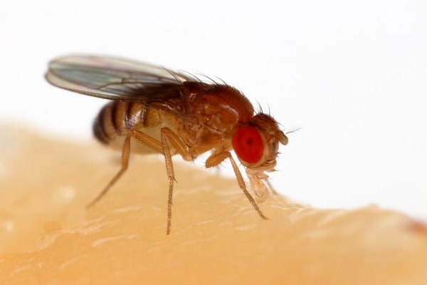
In biology there are a couple of truly crucial model animals and insects. Not that they’re particularly good students, or pick up their own trash, but in the sense that they have become standard model organisms for research. Aside from genetic research, the FlyEM project seeks to fully map a little fly’s brain’s neural connections. This common fly, called drosophila melanogaster (or ‘lesser fruit fly’) has been the subject of a lot of genetic studies, but this study of its brain structure may provide insights in how our brain works as well.
Based on electron microscope images of thin slices of a drosophila brain, the three-dimensional structure of this tiny brain is reconstructed to not only determine the location of each neuron, but also their connections with other neurons. We know that about two-thirds of their brain is dedicated to processing the visual information from their relatively advanced compound eyes, but a lot is still unknown about how this is done, or how the brain’s structure develops.
If it’s always been your dream to tinker with a little fruit fly’s brain, you can do so yourself using the open source FlyBrainLab tool provided, along with the freely available data sources. This tool does not just allow one to visualize the drosophila brain in great detail, but also to create executable circuits and study their functionality. With neurobiology still a largely unexplored territory, this makes for an amazing tool to make this research accessible to anyone.
(Thanks for the tip, [Hernandi Krammes])


