For [Peter Jansen], the most interesting course in grad school was Advanced Brain Imaging; each class was a lecture followed by a trip to the imaging lab where grad students would take turns being holed up in a MRI machine. A few years into his doctorate, [Peter] found himself in a very opportune situation – his local hackerspace just acquired a shiny new laser cutter, he had some free time on his hands, and the dream of creating a medical imaging device was still in the back of his mind. A few weeks later, the beginnings of an open source CT scanner began to take shape.
This isn’t an MRI machine that [Peter] so fondly remembered from grad school. A good thing, that, as superconducting magnets chilled with liquid helium is a little excessive for a desktop unit. Instead, [Peter] is building a CT scanner, a device that takes multiple x-ray ‘slices’ around an axis of rotation. These slices can then be recompiled into a 3D visualization of the inside of any object.
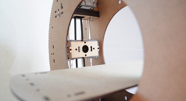
The mechanics of the build are a Stargate-like torus with stepper motor moving back and forth inside the disk. This, combined with the rotation of the disk and moving the bed back and forth allow the imager to position itself anywhere along an object.
For the radioactive detector, [Peter] is using a CCD marketed as a high-energy particle detector by Radiation Watch. Not only does this allow for an easy interface with a microcontroller, it’s also much smaller than big, heavy photomultiplier tubes found in old CT scanners. As for the source, [Peter] is going for very low intensity sources, most likely Barium or Cadmium that will take many minutes to capture a single slice.
The machine operates just above normal background radiation, so while being extremely safe for a desktop CT scanner, it is, however, very slow. This doesn’t bother [Peter], as ‘free’ time on a CT scanner allows for some very interesting, not seen before visualizations, such as a plant growing from a seed, spreading its roots, and breaking the surface as a seedling.
[Peter] still has some work to do on his desktop CT scanner, but once the stepper motor and sensor board are complete, he should be well on his way towards scanning carrots, apples, and just about everything else around his house.


Very cool. I approve.
This can be so awesome with some lead shielding, some collimation, possibly multiple radiation detectors, and a suitable x-rax source. http://www.newtonscientificinc.com/4-watt-monoblock/ However, I could not figure out the pricing for a x-ray source.
Any x-ray source and you are going to need to be licensed by the local govt for the operation of that source, legally at least.
I would expect that this depends strongly on the used intensity?
Im not sure, it may vary from location to location. Possibly the CDRH might be able to tell you.
A roll of sellotape / scotch tape is an x-ray source. No, really – google it! If you unstick an whole roll really quickly it gives off a bunch of different frequency radiation, including x-rays. Not sure if it’s enough to be useful, but stil..
You however need high vacuum in order for it to work, even then the photon energy is something like 15keV (too little for anything usefull)…
The cost of the vacuum setup will be far greater then an actual working x-ray tube (or entire head from a medical device) off ebay…
But only in a high vacuum.
These monoblock sources are in the $1k-3k range.
where are you going to get the radiation source for it to work?
most sources are carefully controlled by the government especially in today’s day in age where terrorists could use the sources to make nuclear bombs
Anyone can buy a test source. There is not enough radioactive material in them to matter in the scheme of things.
You don’t need radioactive materials. X-rays are produced by sufficiently accelerating electrons, then suddenly stopping them. Many common vacuum tubes will produce small amounts of x-rays when driven at higher than intended voltages – sufficient to expose covered photographic film for traditional, 2D x-ray images.
Correct. Ben Krasnow has a video of such a setup:
http://www.youtube.com/watch?v=yLSu_UjrcUA
Easiest way to get a radiation source? Buy a smoke detector.
Smoke detectors use Americium as a source of alpha particles. Alpha is blocked by just about anything – skin, cellophane wrap, and 10 inches of air. It’s not suitable for imaging applications.
You can occasionally find ore samples on eBay with CPMs in the 20K range. I don’t know if that’s high enough for this application, and it’s not directed radiation, as an X-ray tube would be, but it’s something to consider.
An uncapped BJT will detect all kinds of radiation from IR down to cosmic rays. I use 2N5055 and 2N2222 with appropriate circuitry. Grind off the cap, shield it from things you don’t want, and amplify voltage by about 10^6 to get clean pulses. (I really should make a video about that.) The 2N2222 has a small capture aperture, so it might be suitable for this application.
Americium also produces gamma rays and soft X-rays.
The US AEC (before the Energy czars era) used to send free sample alpha sources to anyone who asked. It was a touch of radium paint on the head of a needle. I used one in a DIY cloud chamber in high school and it provided nice traces.
I’ll just leave this here: http://www.dangerouslaboratories.org/radscout.html
CRT.
To my knowledge the radiation-watch sensor is nothing but a CCD that is shielded against visible light. Now how about using a 2d-CCD sensor, e.g. from a cheap camera, to acquire a lot of trajectories through the body at once? Darkness would be obligatory, obviously. Could that work?
Back constructing that is a piece of cake, inverse radon transformation with filtering is quite simple.
Yes that works (certain constraints apply). A colleague and me have done it. I can probably post a link to a paper, if there’s interest.
Yes please.
http://dx.doi.org/10.1109/NSSMIC.2009.5402242
Sorry – it’s not freely available, if you don’t have access to the IEEE digital library yourself, try finding someone at a university, most technical faculties will probably have access.
If you have any specific questions, post them here, I’ll try to answer them.
Lovely idea, I’m currently working on x-ray crystallography using a microbeam soft x-ray tube http://www.hardhack.org.au/crystallography and am in the process of prototyping a fine apature radiation detector using Si Pin photodiodes. I was intending rotating the crystal and building up a raster scan image of the refraction energy. Not ready to publish, yet but getting very close to a working detector.
The tube looks really neat, where (and for how much) did you get it?
I still feel sceptic about x-rays, I wouldn’t use it in my bedroom…
Not reading the linked article is understandable, but did you even read the description? Your kind of baseless paranoia is one of the things holding back technological advancement in radiology. The amount of radiation put off by this device is so small, it’s difficult to even measure, much less cause a problem with your health.
http://en.wikipedia.org/wiki/Magnetic_resonance_imaging
Radiation here at anything. Research is conducted by means of a magnetic field, and measurement of change of this field depending on a resonance which arises at its interaction with cores of atoms. The electromagnetic field too can make not so good impact on a human body, but it it is impossible even will compare to to ionizing radiation influence. Simply all are frightened by the word nuclear in the technique name because not all know that it there means.
The project in the post is NOT an MRI, which wouldn’t put out ionizing radiation. The project is a CT scanner, which DOES use ionizing x-rays.
If the detector needs a certain number of photons to get a good exposure – does it matter whether it’s a short dosage of a high intensity source, or a long dosage from a low intensity source ?
The first option is more desireable, with the later you will also integrate the electronic noise of the detector, resulting in a lower SNR.
Great project!
Superconducting magnets aren’t required for MRI. I stumbled across an educational MRI kit that uses the Earth’s magnetic field instead. http://www.magritek.com/products-terranova-overview An image takes hours to capture and is very low res, but it’s still pretty cool. They also make a permanent magnet MRI system that captures very detailed images of small objects.
Whoa! That’s seriously cool. I know there are MRI systems that use super powerful specialty permanent magnets, but I didn’t think it was possible to acquire any sort of image using Earth’s magnetic field.
The lack of pricing on that kit you linked is discouraging, however….
The magnetic field causes certain atomic nuclei to spin and precess like a top wobbling. The nuclei can’t line up perfectly with the magnetic field because that would violate the uncertainty principle, so it has to point at some angle from the external field. It spins around this field with a speed of rotation which is a function of the strength of the field.
While spinning, if you pulse an RF signal at the frequency of rotation, you can get the nuclei to flip end-for-end. This absorbs energy from the RF pulse (which can be measured), and at some future time the nuclei will flip back and emit an RF photon (which can also be measured).
The preceeding will be true for any strength magnetic field. The medical scanners use high strength fields so that they can use higher frequency RF photons, which have correspondingly shorter wavelengths which gives them more resolution. If you can live with lower resolution then you can use a weaker field.
The medical scanners use very large magnets because the field has to be uniform. Magnetic fields diverge outside of the torus, so the nuclei in those sections see a different field strength, rotate at a different frequency, and contribute nothing to the scan. If the field isn’t uniform within the patient, then those sections won’t contribute.
It’s my understanding that field uniformity is the biggest issue in MRI design. If all you want to do is “see the effect”, you can suspend a test tube containing your sample between the poles of a “C” magnet with a field coil to generate the RF. Google “NMR” instead of “MRI”, the name was changed because “nuclear” sounds scary.
You can just build your own source like this guy did
http://hackaday.com/2013/06/02/building-a-miniature-x-ray-tube/
or go on http://www.unitednuclear.com/ and get whatever radiation source you like as long as you are in the U.S.
Brilliant project. I wish I had the time and knowledge to build something like this.
I remember reading about (mit?) doing research on low power MRI few years ago. There was even talk or being able to build cheap portable MRI machines that use cellphones for computing and display. It was supposed to be possible thanks to new computational techniques (compressed sensing?)
This one?
http://martinos.org/lfi/pdf/LFI_JMR_published.pdf
Big difference between MRI and CT is the use of X-rays. X-rays cause cancer. Currently the chance of dying because of a radiation induced malignity is estimated at 5% per Sievert. That’s a lot of dose, but on the other hand low doses spread over a large population are not insignificant. that’s why medical and industrial applications of ionizing radiation are heavily regulated. For example a medical CT scan can result in to up to a few mSv, so about 1 in 50000 CT scans lead to a terminal cancer (if the patient lives long enough to develop it, typically 25 years after exposure).
Don’t expect a rapid growth of unregulated X-ray use ;)
The low dose rate radiation effects are statistically so serious that they have to be extrapolated from higher dose rates. The history of the linear non-threshold model (not taking in account the cells’ self-repair mechanisms) that’s the basis of contemporary legislation and assumptions is dated to the politically motivated battles around the ban on nuclear testing. Too bad the fallout of this may be worse than the fallout from the actual tests…
Very impressive!
However, he mixed up one aspect – the Radon transform is not directly going to reconstruct his images. Radon is the integral along the lines in the 2D image. Reconstruction requires the “inverse” thereof which does not exist. Typically, a filtered backprojection is used as a good approximation.
Find a copy of the dead tree book “The Scientific American Book of Projects for the Amateur Scientist” by C.L.Stong. It shows how to make a working X-Ray tube and other safe and not-so-safe projects. I built the NMR and a Low Speed Wind Tunnel from it (or a derivitive) when I was in high school (40 years ago). Still a good book for reference on the pre-micro computer age. In whatever you do, be careful out there.
Looked into using a modified Cool-X to do this a while back, trouble is as ionising sources even broken ones are restricted.
I think NASA use them though, maybe someone can source one?
Alternate ideas, some Russian tubes (shunt regulators especially) are nice X-ray sources and a very minor tweak yields a collimated beam suitable for DIY CT use.
disclaimer:- yadayada etc
For the ‘danger hearted’, pull out an old archived copy of ‘The Scientific American Book of Projects for the Amatuer Scientist’ by C.L.Stong. It has some great projects in it, anything from seismic sensors, x-ray and NMR machines that work, robot & computers powered by relay logic, Lots of fun stuff, and all ultimately doable in a well equipped home lab.
It is not easy to find, and the electronic versions I have seen are missing some of the ‘dangerous’ experiments. … But those are the fun ones to learn about even if you don’t do them.
(In High School I built the NMR Machine, low speed wind tunnel, and a couple of other projects. But that was back in the day when Knights were Bold and it was OK to do ‘dangerous’ things. BTW, the stuff I did wasn’t dangerous. :-) )