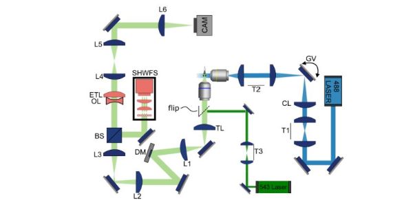

Key to understanding something like epilepsy is to be able to record highly transient events in biological tissues. Generally this is done using light sheet microscopy, which provides effectively a 2D ‘slice’ of the tissue in question, but to observe a brief event in a larger biological system you need to be able to rapidly change the layer and focus between the virtual layers. This is what [Bingxi Liu] et al. al did using adaptive optics with an electrically tunable lens (ETL) in order to capture seizures in the brain of zebrafishes.
Their system can capture a volume of 499 × 499 × 150 μm3 at 4 volumes per second, which is large enough to fit optically transparent zebrafish larva into. The optical setup is shown in the above image, with the design based on the OpenSPIM platform for selective plane illumination microscopy.
Here the 488 nm laser provides the illumination (excitation) of the layer, while the 543 nm laser is for calibration purposes. The ETL is thus in the imaging path that allows for capturing by a digital camera, while a beam splitter directs part of the captured data to a Shack-Hartmann wave front sensor (SHWFS), which is part of the adaptive optics system.
After a seizure was induced in the zebrafish larva using the drug pentylenetetrazol the results were recorded using this system. It showed the seizure’s origin in the posterior brain, with subsequent propagation to the anterior before subsiding gradually over tens of seconds.
This system should be quite useful even outside of seizure research, as there are a lot of 3D systems in biology where having a relatively high-speed microscopic capture can be very revealing.