Who doesn’t want an X-ray machine? But you need a special tube and super high voltage, right? [Project 326] says no, and produces a USB-powered device that uses a tube you can pick up two for a dollar. You might guess the machine doesn’t generate X-rays with a lot of energy, and you’d be right. But you can make up for it with long exposure times. Check out the video below, with host [Posh Arthur].
The video admits there are limitations, of course. We were somewhat sad that [Project 326] elected not to share the exact parts list and 3D printed files because in the unlikely event someone managed to hurt themselves with it, there could be a hysterical reaction. We agreed, though, that if you are smart enough to handle this, you’ll be smart enough to figure out how to duplicate it — it doesn’t look that hard, and there are plenty of not-so-subtle clues in the video.
The video points out that you can buy used X-ray tube for about $100, but then you need a 70kV power supply. A 1Z11 tube diode has the same basic internal structure, but isn’t optimized for the purpose. But it does emit X-rays as a natural byproduct of its operation, especially with filament voltage.
The high voltage supply needs to supply at least 1mA at about 20 kV. Part of the problem is that with low X-ray emission, you’ll need long exposure times and, thus, a power supply needs to be able to operate for an extended period. We wondered if you could reduce the duty cycle, which might make the exposure time even longer, but should be easier on the power supply.
The device features a wired remote, allowing for a slight distance between the user and the hot tube. USB power is supplied through a USB-C PD device, which provides a higher voltage. In this case, the project utilizes 20V, which is distributed to two DC-DC converters: one to supply the high-voltage anode and another to drive the filament.
To get the image, he’s using self-developing X-ray film made for dental use. It is relatively sensitive and inexpensive (about a dollar a shot). There are also some lead blocks to reduce stray X-ray emission. Many commercial machines are completely enclosed and we think you could do that with this one, if you wanted to.
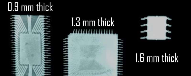
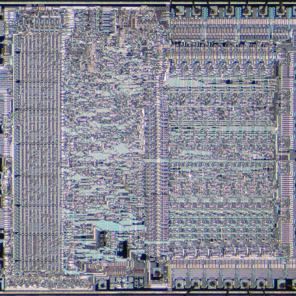
You need something that will lie flat on the film. How long did it take? A leaf image needed a 50-minute exposure. Some small ICs took 16 hours! Good thing the film is cheap because you have to experiment to get the exposure correct.
This really makes us want to puzzle out the design and build one, too. If you do, please be careful. This project has a lot to not recommend it: high voltage, X-rays, and lead. If you laugh at danger and want a proper machine, you can build one of those, too.


Rustin Cohle wearing a surgeon’s hat and dragging on a ciggy definitely inspires confidence in this endeavor, good choice.
Driving the anode voltage out of USBC for extended periods is worth looking into by itself. All kinds of fun stuff you could do with that… most of them shady, I like it
What’s the difference between “filament voltage” and “high voltage”? It sounds like you’re saying both are involved.
Filament voltage drives the tube’s heater (literally a tungsten filament like a light bulb), for this tube it’s 1.2V at probably 200 mA or so. This emits electrons, as the cathode.
The high voltage (10s of kV, tiny currents in this case) accelerates the electrons, which hit the anode and generate X-rays.
Thank you, that makes it clear. Here’s where I misunderstood: “But it does emit X-rays as a natural byproduct of its operation, especially with filament voltage.”
The tube will produce a bit of output even without heating the filament. Apply a bit of voltage to the filament to heat it, and the x-ray output will increase dramatically.
But, yeah, (sorry Al) it’s a little unclear as written.
This is officially the most terrifying Hackaday headline of the past year.
They’re basically taking advantage of the existing tiny x-ray output of a standard rectifier tube, and then using exposure times up to ten hours long at an extremely close distance. The emitted photons are completely blocked by even the thin copper layer of a PCB.
So while you don’t want to put your finger directly on it, you’re probably as safe a few meters away as if you were near an old tube TV.
Yeah, but if you sat too close to the tele, your eyes would go square – that’s what my mum used to tell me…
Just spend another decade in front of the boob-tube at +/- 45* and you’ll be indistinguishable from normal.
I love the video, it is true, generating X-rays is easy enough it is not like it is some really complex process (X-rays were discovered by Wilhelm Conrad Röntgen in 1895). The main thing that has changed over the last 130 years years is our knowledge of how really bad they are for human health. Soft in some ways are even more scary than hard because they are so easily absorbed by soft tissue.
True, low energy “soft” X-rays deposit almost twice as much dose in the first millimeter or two. Scary indeed. The more penetrating higher energy “hard” x-rays get absorbed less in the skin, and go right on through to deposit dose in internal organs.
Sticking your fingertip on the output window of a low-energy x-ray tube is a dumb idea, not because low-energy x-rays deposit more dose, but because you’re sticking your finger a couple of millimeters away from a point radiation source, so the dose per square millimeter is many thousands of times higher than it would be in a usual x-ray imaging setup.
If you can figure a digital sensor for the X-rays, toggling the tube on and off could increase resolution and reduce noise though lock-in sensing. Similar to https://hackaday.com/2024/10/28/lock-in-thermography-on-a-cheap-ir-camera/
Uh, no, not in most reasonable situations.
If you have high contamination (noise) from other radiation sources and need to isolate the signal from your desired source then, yes, lock-in sensing can help.
But in x-ray imaging you don’t usually have a high background radiation rate to contend with.
I think you misunderstood lock-in sensing. The idea is to correlate the signal from your sensor with a signal you have control of in the device under test.
You typically don’t have control on a millisecond time scale of the density of an item you want to x-ray.
Do they sell 3d printer filament that can shield gamma rays?
Nobody is talking about gamma rays here.
But any material will absorb (“shield”) gamma rays. Denser materials allow you to make the absorber thinner for a given amount of shielding. To be more specific, you’ll need to know your gamma energy spectrum and the characteristics of the material.
Interestingly, kilogram for kilogram, plain old air makes a wonderful shield, and has the neat property that its shielding goes as the square of the thickness used, where other materials’ shielding vs thickness are closer to linear. For a given cross section area, 10 meters of air weighs less than a millimeter of lead but usually provides much better dose reduction.
That “square of the thickness” relationship is just the inverse-square law that governs free space loss, and it’s due to geometry, not the medium. You multiply that loss by the inverse-exponential attenuation that does depend on the medium but as the name suggests has diminishing effects as the shielding gets thicker.
So it makes sense to use a combination of a shielding enclosure and distance from it.
OK, right — If someone has to explain the joke, it’s not funny.
Yes. https://www.prusa3d.com/product/prusament-petg-tungsten-75-1kg/
Interesting stuff to be sure, but be aware that although the “75% tungsten” (by weight) claim might be correct, that means that only one sixth of the volume is tungsten: the rest is PETG binder. If you’re looking for bulk shielding material, and don’t need the custom shapes afforded by 3D printing, even tin foil will perform as well. Plain old 63:37 solder melted into your desired mold shape will be much better.
And if you don’t want to deal with molten metal, tungsten powder is pretty cheap, and epoxy makes a fine binder.
“soft X-rays – Not as safe as it sounds. I’d rather play with my dental X-ray head and get an exposure in a few ms instead.
soft X rays — not as dangerous as the fearmongers would have you believe.
But, yes, still dumb to play with without appropriate safeties and knowledge.
As soft as skin cancer and cateracts.
If you mean cataracts, yes, ionizing radiation has been identified as a risk factor, just like sunlight. Soft x-rays aren’t unique.
In case it’s not clear: Yes, it’s not good to go dosing yourself with ionizing radiation at any level more than necessary. But soft x-rays aren’t especially worse than any other wavelength.
They’re nasty in uses like x-ray crystallography as they are a beam and the whole energy lands on one spot.
That spot goes necrotic and the gangrene kicks in.
Yeah… don’t do that. Especially if it worked like that.
The dose makes the poison, doesn’t it?
At 20 kv you’ll mostly produce x-rays at a 2-4 kv energy, a very soft beam that could be absorbed by even an inch or two of water.
With a lot of time you might get a very detailed mamogram of a fruit, but you’d have to image for ages to do much with taxidermy.
At 20 kV accelerating potential you might produce x-ray photons mostly in the sub-5keV range, but approximately zero of those will escape the anode and the glass envelope. Any that will would be mostly absorbed in much less than a millimeter of water ( a few dozen microns on average).
The average energy of photons leaving a 20 kV source through a glass envelope will be more like 15 keV. About 1.5% of those will make it through an inch of water or fruit or body tissue — plenty to make an image.
I bet there are some old cobbler fluoroscopes out there somewhere…
An x-ray lens
https://phys.org/news/2025-06-wafer-lens-ray-size.html
More:
https://phys.org/news/2025-06-high-explosives-motion-molecules-chemical.html
https://phys.org/news/2025-06-composite-materials-broad-spectrum-synergistic.html
United Nuclear (https://unitednuclear.com/search_result.html?main_page=search_result&search_in_description=1&keyword=X-ray) is an online retailer that sells everything you need to experiment with “soft X-rays” except for film, including a compatible HV power supply, lead shielding to keep you safe, and rectifier tubes that they have all been tested to make sure they actually emit X-rays. (I have no connection to the company).
BTW, most dentists have converted to digital X-ray sensors, so eventually it will be either hard or expensive to buy X-ray film.
I wonder how sensitive “full frame” DSLR sensor are to low energy X-rays.
A DSLR sensor can work to detect x-rays. Sort of.
The problem is that the sensitive thickness in a silicon imaging chip is tiny — a few microns. And silicon is a low atomic number so its absorption (and therefor detection) of x-rays per unit volume is quite low.
Most x-rays sail on through, undetected, unless the x-ray energy is low enough to be stopped by the relatively poor and thin absorber: remember, silicon imagers are designed to absorb and detect photons around 1-2 eV of energy. It’s as transparent as glass to typical x-ray energies in the 50 keV range.
But if you use x-ray energies below 10 keV you get usable detectability. Unfortunately, the cover glass on the imaging chip itself blocks most of that.
So if you want to use an unmodified DSLR to detect x-rays, you’re stuck using energies high enough to get through the glass, but low enough for a decent fraction of the photons to be absorbed by the silicon: between 10-20 keV.
You also need to exposure time to be short enough so your image is not dominated by dark noise (or light leakage through the viewfinder). A mammography tube running with no filtration at 28 kV and a few mA works – at 20 cm you can use exposure times of one second to get a reasonable image. Don’t do this on living human tissue: that’s an irresponsible amount of skin dose.
But, basically it’s a dumb idea: You’re almost always better off to use a scintillator screen and a good fast lens, unless you really want high resolution images at 5-10 keV and can take the window off the chip.