Neuroscientists have been mapping and recreating the nervous systems and brains of various animals since the microscope was invented, and have even been able to map out entire brain structures thanks to other imaging techniques with perhaps the most famous example being the 302-neuron brain of a roundworm. Studies like these advanced neuroscience considerably but even better imaging technology is needed to study more advanced neural structures like those found in a mouse or human, and this advanced MRI machine may be just the thing to help gain better understandings of these structures.
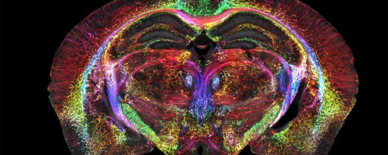
A research team led by Duke University developed this new MRI technology using an incredibly powerful 9.4 Tesla magnet and specialized gradient coils, leading to an image resolution an impressive six orders of magnitude higher than a typical MRI. The voxels in the image measure at only 5 microns compared to the millimeter-level resolution available on modern MRI machines, which can reveal microscopic details within brain tissues that were previously unattainable. This breakthrough in MRI resolution has the potential to significantly advance understanding of the neural networks found in humans by first studying neural structures in mice at this unprecedented detail.
The researchers are hopeful that this higher-powered MRI microscope will lead to new insights and translate directly into advancements healthcare, and presuming that it can be replicated, used on humans safely, and becomes affordable, we would expect it to find its way into medical centers as soon as possible. Not only that, but research into neuroscience has plenty of applications outside of healthcare too, like the aforementioned 302-neuron brain of the Caenorhabditis elegans roundworm which has been put to work in various robotics platforms to great effect.


there’s 10.5 tesla and 11.7 tesla and more just google
23.5T?
https://archives.cnrs.fr/presse/article/1689?&debut=8
in 2009 that was…
That’s for NMR which is related but not the same as MRI
Isn’t 6 orders of magnitude from 5 microns = 5 m?
It might just be two per dimension, since MRI’re 3D. 100*100*100 = 1 million.
Maybe, but “6 orders of magnitude” is more catchy and clicbaity.
From comments on Hackernews so far this technique only works on very small dead things so maybe not great for humans just yet.
Thanks for that–saw an article about this but it didn’t say whether it could be used on living material. This would be comparable to a low-grade 3D electron microscope in my opinion. It’s still a cool accomplishment, just not what everyone thinks of as a MRI.
It looks like this is a tool for basic medical research, not for clinical medicine.
Or forensic clinical medicine. Good luck finding tissues / organism that does not move at this scale. One would need to be deep-frozen before getting a high-res MRI at the current speed.
That makes a lot more sense. With announcements like this, there’s always a catch.
That’s great. But does it require some of the vanishing supply of liquid helium to function?
Use caution in viewing the link. The Duke imaging video has ‘music’ meant to cause brain damage for later examination.
Well, would you look at that — my old stomping grounds showing up on Hackaday. :)
I worked at CIVM from about 2002 to 2010. I was just a developer/analyst, so I didn’t get to build or run the equipment. I did get an office with tanks of liquid nitrogen and liquid helium just outside the door. (I got an office with a DOOR!)
It was the coolest place I’ve ever worked, bar none. I miss it a lot. At that time, we already had the 9.4T magnet, along with a 7T and a 2T. Our best images out of the 9T at that point were I think 20um resolution. They had plans for getting to 10um, but I thought 5um was pretty much physically impossible.
To get more resolution, you need some combination of higher field strength, better SNR in the transmitters/coils/receivers, and longer scan time.
Higher field strengths are an issue, because that increases the frequency you need to transmit and receive, and when those frequencies get high enough they don’t penetrate far enough into tissue. 9T is fine for a rodent brain, but to use it with people, I think you’d need internal/implanted coils, unless the stuff you want to image is within a few cm of the surface. (Which kind of goes against the whole “non-invasive” part.)
To improve SNR, they were working with superconducting receiver coils; that’s tricky, because they need to be close to the subject, which typically does NOT want to be at cryo temps. I haven’t yet read the paper to see if they’re using superconducting coils here. (The field magnets were all superconductive.)
Scan time was already running to many, many hours. I’m guessing this scan took more hours than that. For in-vivo imaging, that gets to be a problem. For in-deado, you have the advantage of a subject that will keep VERY still indeed, and not need breaks for bathroom. Or breathing, or heartbeats — really high-resolution work in-vivo was gated against both respiration and heartbeat. They’ve got beautiful 3D movies of beating rat hearts imaged in live animals.
Man, I miss that place…
Too bad it isn’t real-time..