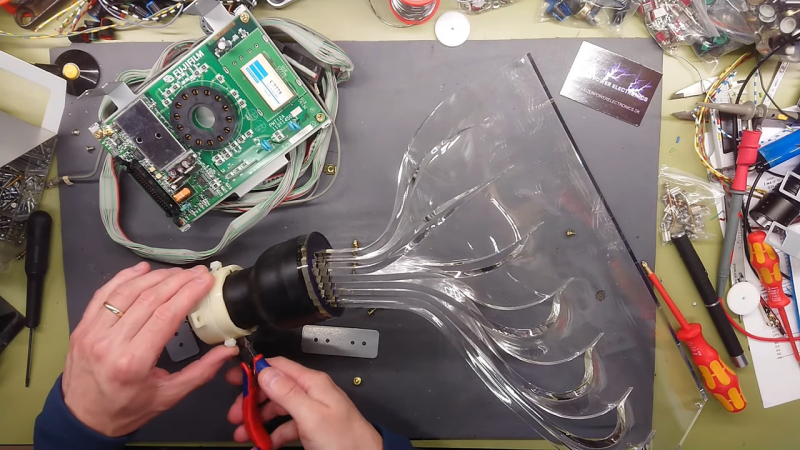
We’ll just go ahead and say it right up front: we love teardowns. Ripping into old gear and seeing how engineers solved problems — or didn’t — is endlessly fascinating, even for everyday devices like printers and radios. But where teardowns really get interesting is when the target is something so odd and so specialized that you wouldn’t normally expect to get a peek at the outside, let alone tramp through its guts.
[Mads Barnkob] happened upon one such item, a Fujifilm FCR XG-1 digital radiography scanner. The once expensive and still very heavy piece of medical equipment was sort of a “digital film system” that a practitioner could use to replace the old-fashioned silver-based films used in radiography, without going all-in on a completely new digital X-ray suite. It’s a complex piece of equipment, the engineering of which yields a lot of extremely interesting details.
The video below is the third part of [Mads]’ series, where he zeroes in on the object of his desire: the machine’s photomultiplier tube. The stuff that surrounds the tube, though, is the real star, at least to us; that bent acrylic light pipe alone is worth the price of admission. Previous videos focused on the laser scanner unit inside the machine, as well as the mechatronics needed to transport the imaging plates and scan them. The video below also shows experiments with the PM tube, which when coupled with a block of scintillating plastic worked as a great radiation detector.
We’ve covered a bit about the making of X-rays before, and a few of the sensors used to detect them too. We’ve also featured a few interesting X-ray looks inside of tech, from a Starlink dish to knock-off adapters.
Thanks for the heads up on this one, [Adrian]!


Thank you very much for featuring some of my work :)
I thought alpha particles were blocked by an inch of air, and also a sheet of paper or a sheet of foil.
How are you detecting them in this setup? If the foil is open at the end, how are you eliminating ambient light?
The foil is wrapped around several times, there is no ambient light getting in. A user on youtube commented that while Thoriated welding rods has a whole breakdown range of radiation that ends up with some weak Gamma. Because as I write and you say, Alpha and Beta should be blocked by the materials in place.
don’t understand this, how does such a low resultion light tube yield high resolution x-rays??
Frickin’ lasers, man.
The magic is in the plate the image is recorded on: it’s a stimulated emission phosphor: the x-ray exposure stores a latent image in the phosphor. When struck by a scanned (red) laser beam, the phosphor emits blue light proportional to the x-ray energy absorbed at that spot.
So the red laser is scanned right in front of that linear pickup. The PMT is blind to red light, but sees the blue photons liberated by the scanned laser spot. The plate is advanced through the reader line by line. The resolution is determined by the size of that laser spot (and the bandwidth of the PMT and the raster pitch…)
Dan could tell you all about the business of handling these things, I’m sure.
And, BTW, the residual image left over after reading must be erased from the plate before the next x-ray exposure. Done by a big frickin’ white light. The plate is resusable pretty much indefinitely, or until it gets handled to death.
Phosphor must have a long decay time with that setup.
The phosphor doesn’t decay. The latent x-ray image is stored indefinitely(*) until it is stimulated to emit its blue photons by tickling the phosphor with red light. That’s the magic of the thing.
( (*) though the latent image will very slowly fade away eventually. You want to read out the plate promptly after exposure – certainly the same day.)
It is called Image Reconstruction from Projections and involves a lot of math and computing. Like constructing a 3D image from a series of shadows. It looks like the light guide on the tube in the original photos detects x-rays in a thin strip as it rotates around a target. This might be OK https://www.youtube.com/watch?v=gEQt5HacXJc
No, not at all. You’re describing tomography, a completely different thing. This uses no math, no computing. The image in the phosphor is just the image that gets read out. In principle you don’t need a computer, except in practice the output is digitized, stored, and displayed digitally.
Ah, I misunderstood. You mean the original plate scanner? Yes. I thought the OP was heading towards building something else.
That acrylic light pipe is *brilliant*.