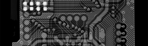
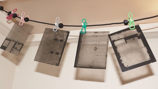
Digital X-Ray Scanner Teardown Yields Bounty Of Engineering Goodies

We’ll just go ahead and say it right up front: we love teardowns. Ripping into old gear and seeing how engineers solved problems — or didn’t — is endlessly fascinating, even for everyday devices like printers and radios. But where teardowns really get interesting is when the target is something so odd and so specialized that you wouldn’t normally expect to get a peek at the outside, let alone tramp through its guts.

[Mads Barnkob] happened upon one such item, a Fujifilm FCR XG-1 digital radiography scanner. The once expensive and still very heavy piece of medical equipment was sort of a “digital film system” that a practitioner could use to replace the old-fashioned silver-based films used in radiography, without going all-in on a completely new digital X-ray suite. It’s a complex piece of equipment, the engineering of which yields a lot of extremely interesting details.

The video below is the third part of [Mads]’ series, where he zeroes in on the object of his desire: the machine’s photomultiplier tube. The stuff that surrounds the tube, though, is the real star, at least to us; that bent acrylic light pipe alone is worth the price of admission. Previous videos focused on the laser scanner unit inside the machine, as well as the mechatronics needed to transport the imaging plates and scan them. The video below also shows experiments with the PM tube, which when coupled with a block of scintillating plastic worked as a great radiation detector.

We’ve covered a bit about the making of X-rays before, and a few of the sensors used to detect them too. We’ve also featured a few interesting X-ray looks inside of tech, from a Starlink dish to knock-off adapters.

Continue reading “Digital X-Ray Scanner Teardown Yields Bounty Of Engineering Goodies”

A Perfect Rose, A Stepper, An X-Ray Machine, And Thee

The Day of Compulsory Romance is once more upon us, and in recognition of that fact we submit for your approval an alternative look at one of the symbols of romantic love: an X-ray of a rose.

Normally, diagnostic X-rays are somewhat bland representations of differential densities of the tissues that compose variousĀ organs and organ systems, generally rendered in shades of gray. ButĀ [Linas K], a physicist with time to kill and access to some cool tools, captured the images in the video below not only in vivid color, but as a movie. The imaging side of the project consists of a low-power X-ray tube normally used for non-clinical applications and a CMOS sensor panel. The second video shows that [Linas] did an impressive upgrade on the X-ray controller, rolling his own from an FPGA. This allowed him to tie in a stepper motor to rotate the rose incrementally while taking images which he stitched together in post.

Watching the interior structure of the flower as it spins is fascinating and romantic in its own right, for certain subsets of romance. And really, who wouldn’t appreciate the work that went into this? But if you don’t have access to X-ray gear, fear not — a lovely Valentine’s gift is only a bottle of ferric chloride away.

Continue reading “A Perfect Rose, A Stepper, An X-Ray Machine, And Thee”

Hackaday Prize Semifinalist: Low Cost Radiography

For the past year, [Adam] has been working full-time on developing a low-cost x-ray system for developing nations. He has more than 3,500 hours into the project. A few months ago, we announced the 2015 Hackaday Prize, with a theme of, ‘build something that matters.’ A low-cost x-ray would certainly matter to the two-thirds of the world’s population that does not have access to medical radiography, making this project a great entry for The Hackaday Prize.

[Adam]’s portable x-ray system consists of an x-ray tube encased in an epoxied, 3D printed enclosure filled with dialectric oil. This tube is tucked away inside a beautiful case with just a single 12VDC input and an easy to understand user manual. This is just very high voltages and x-rays, nothing [Adam] hasn’t handled (safely) before. The real trick is in the imaging, and for this, [Adam] is using a phosphor screen to turn that x-ray exposure into something visible, an off the shelf x-ray sensor, and a prism to adapt the sensor to the phosphor screen.

The results are incredible. After taking a few pictures of what he had on hand, [Adam] can see the bond wires inside the microprocessor of a calculator. That’s more than sufficient for medical imaging – the goal of the project – and cheap enough to send it to the far-flung reaches of the planet.

The 2015 Hackaday Prize is sponsored by:

Continue reading “Hackaday Prize Semifinalist: Low Cost Radiography”