Fluoroscopy is probably the best-known method of X-ray imaging: an X-ray beam passes through the subject to be imaged, and the transmitted X-rays illuminate a phosphor screen. Dense objects, such as metal or bone, cast a shadow on the screen, which provides a real-time image of the subject’s interior. Already having access to X-ray sources, [MarcellF]’s next step was to investigate common phosphor materials, then synthesize his own.
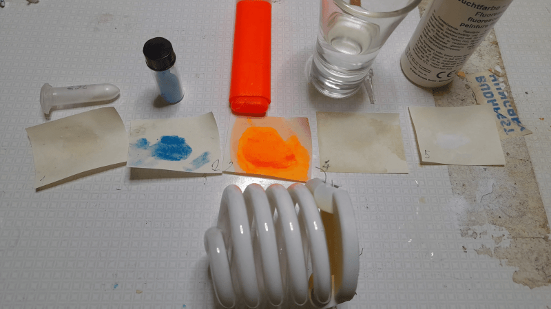
Most common materials that fluoresce under ultraviolet light showed no activity under X-rays: fluorescein, quinine, UV fluorescent paint, and common fluorescent minerals emitted no noticeable glow under 80 kV X-ray stimulation. However, strontium aluminate phosphors did fluoresce well, with a strong afterglow, as did the phosphors in a fluorescent light bulb, some LEDs, and an electroluminescent panel. The electroluminescent panel, which used a zinc sulfide phosphor, was almost as bright as the gadolinium oxysulfide screen from a CT scanner’s detector and had no noticeable afterglow.
One well-known X-ray phosphor is scheelite (calcium tungstate), which [MarcellF] next synthesized. He had previously tested a sample of natural scheelite without success, probably due to impurities. The first step of the synthesis was to melt together potassium nitrate and sodium carbonate, in which [MarcellF] dissolved broken pieces of a tungsten TIG welding rod. This formed sodium and potassium tungstates, which were dissolved and reacted with a calcium chloride solution. This precipitated calcium tungstate, which [MarcellF] annealed to make fluorescent. This produced a blue glow under X-ray stimulation, and doping with lead atoms made it significantly brighter.
We’ve covered several methods of X-ray detection before; most modern fluoroscopes now use a phosphor screen in conjunction with a camera, or sometimes with a photomultiplier tube.


With the availability of optical clear YAG:Ce cut as gemstone “canary yellow yag”, the same stuff is used in “white LED” phosphor for it’s yellow fluorescence, one would expect it to work well quite for this?
A tiny gemstone glued onto a photodiode? The fluorescence lifetime of YAG:Ce is supposedly quite fast (ns range).
I have shot white LEDs with x-rays. They do light up, but only with a tiny fraction of the intensity of a gadox screen.
I have also measured the fluorescence lifetime of the yellow phosphor to be about 1 microsecond. (Measured with a 35 MHz bandwidth detector, so that’s real.)
I’d suspect the longer fluorescence lifetime is from the other stuff in the white phosphor mix. Different materials are optimal for different use cases, but some are more accessible (and less toxic) than others. The gemstone will be only YAG:Ce (and as single crystal), so higher density and more bulk (thickness) than the white LED.
The yellow phosphor component has the 1 us decay time. The red phosphor component is about 2 us. The components are easy to measure with optical filters to separate them. The blue excitation LED light follows the excitation current waveform (<0.1 us in my case, and about the bandwidth of the detector I used).
I don’t know what alchemy is going on to make the yellow so much longer than the pure YAG:Ce decay time (a few dozen ns).
It’s surprising the X-rays didn’t ionize the Hg vapor in the lamp, which should have caused all phosphors to glow. Perhaps it is a low-Hg lamp, with most captured in an amalgam near the filaments?
Further experiment: An LED, though not efficient as a phosphor, can serve as it’s own photodetector. You might add the DC (or pulsating DC) voltage reading across the diode. Also, compare white LED output, with its phosphor, with others, such as blue or red, which might still generate an output by direct conversion of X-ray photons.
X rays would and do ionize the mercury vapor, but at less than 1 Pa of pressure there’s precious little mercury vapor present to absorb the X rays., and only a tiny fraction of that would be ionized, and only a small fraction of the x-ray photon energy would go into the ionization that would produce the UV to excite the phosphor. And there just isn’t that much x-ray photon power to excite it either (milliwatts), so the absolute brightness would be very low.
There is also hundreds of times the mass of phosphor to absorb x-rays (~gram) than there is mercury (milligrams). So you’re left with the bulk of the absorption by the phosphors, and they aren’t terribly good at converting x-ray photons to visible light.
Contrast that with electric arc excitation: basically all the mercury vapor is ionized, with watts of power and producing UV directly, and the tube is warm so the vapor pressure is higher so there’s more mercury present.
Astronauts going into deep space should bring along a zinc sulfide phosphor panel.
Always nice to have a visual indicator.
It should be in the form of glow-in-the-dark letters stating: “If you can read this, you’ll be dead soon.” A radiation exposure that can light up a phosphor enough to see with naked eyes will be lethal inside a day.
I have a sign written in yellow highlighter in my resin printer room: “If you can read this, the UV light is on.” It’s invisible in the normal (yellow) room lighting, but bright when the UV light is on.
The UV light is to neutralize any spills or droplets and incidentally makes them visible to clean up, because the resins fluoresce too. The UV light itself is not visible if the room light is on, and leaving a container of resin exposed if the UV light is on will ruin the resin, hence the sign.