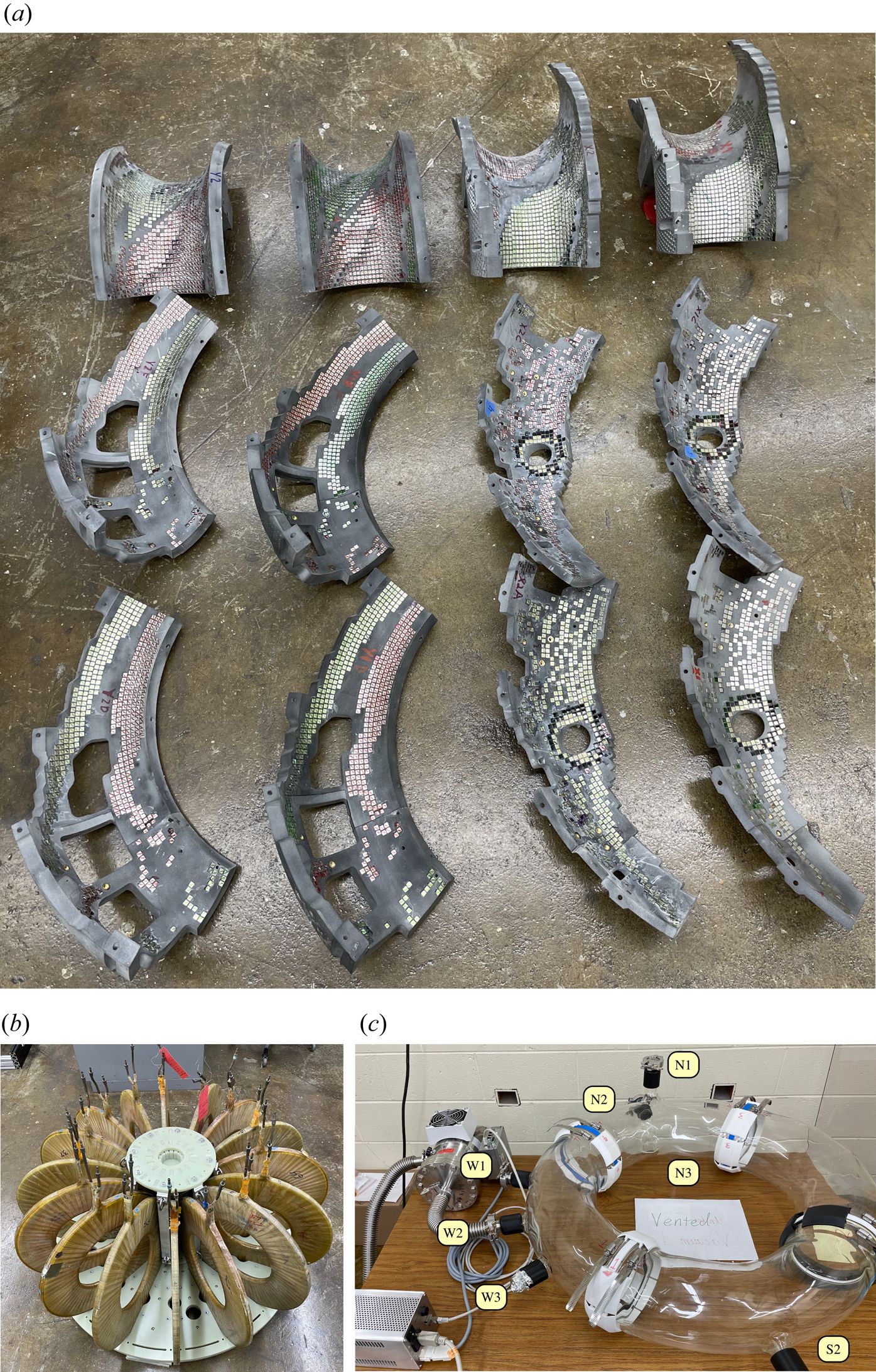
There are probably very few people on this globe who at some point in time haven’t heard the term ‘Higgs Boson’ zip past, along with the term ‘God Particle’. As during the 2010s the scientists at CERN were trying to find evidence for the existence of this scalar boson and with it evidence for the existence of the Higgs field that according to the Standard Model gives mass to gauge bosons like photons, this effort got communicated in the international media and elsewhere in a variety of ways.
Along with this media frenzy, the physicist after whom the Higgs boson was named also gained more fame, despite Peter Higgs already having been a well-known presence in the scientific community for decades by that time until his retirement in 1996. With Peter Higgs’ recent death after a brief illness at the age of 94, we are saying farewell to one of the big names in physics. Even if not a household name like Einstein and Stephen Hawking, the photogenic hunt for the Higgs boson ended up highlighting a story that began in the 1960s with a series of papers.
PhD students spend their time pursuing whatever general paths their supervisor has given them, and if they are lucky, it yields enough solid data to finally write a thesis without tearing their hair out. Sometimes along the way they result in discoveries with immediate application outside academia, and so it was for [Paul Bupe Jr.], whose work resulted in a rather elegant and simple bend sensor.
The original research came when shining light along flexible media, including a piece of transparent 3D printer filament. He noticed that when the filament was bent at a point that it was covered by a piece of electrical tape there was a reduction in transmission, and from this he was able to repeat the effect with a piece of pipe over a narrow air gap in the medium.
Putting these at regular intervals and measuring the transmission for light sent along it, he could then detect a bend. Take three filaments with the air-gap-pipe sensors spaced to form a Gray code, and he could digitally read the location.
He appears to be developing this discovery into a product. We’re not sure which is likely to be more stress, writing up his thesis, or surviving a small start-up, so we wish him luck.
The central selling point of qubit-based quantum processors is that they can supposedly solve certain types of tasks much faster than a classical computer. This comes however with the major complication of quantum computing being ‘noisy’, i.e. affected by outside influences. That this shouldn’t be a hindrance was the point of an article published last year by IBM researchers where they demonstrated a speed-up of a Trotterized time evolution of a 2D transverse-field Ising model on an IBM Eagle 127-qubit quantum processor, even with the error rate of today’s noisy quantum processors. Now, however, [Joseph Tindall] and colleagues have demonstrated with a recently published paper in Physics that they can beat the IBM quantum processor with a classical processor.
In the IBM paper by [Yougseok Kim] and colleagues as published in Nature, the essential take is that despite fault-tolerance heuristics being required with noisy quantum computers, this does not mean that there are no applications for such flawed quantum systems in computing, especially when scaling and speeding up quantum processors. In this particular experiment it concerns an Ising model, a statistical mechanical model, which has many applications in physics, neuroscience, etc., based around phase transitions.
Unlike the simulation running on the IBM system, the classical simulation only has to run once to get accurate results, which along with other optimizations still gives classical systems the lead. Until we develop quantum processors with built-in error-tolerance, of course.
Using technologies like electron microscopy (EM) it is possible to capture molecular mechanisms in great detail, but not when these mechanisms are currently moving. The field of cryomicroscopy circumvents this limitation by freezing said mechanism in place using cryogenic fluids. Although initially X-ray crystallography was commonly used, the much more versatile EM is now the standard approach in the form of cryo-EM, with recent advances giving us unprecedented looks at the mechanisms that quite literally make our bodies move.
Myosin-5 working stroke and walking on F-actin. (Credit: Klebl et al., 2024)
The past years has seen many refinements in cryo-EM, with previously quite manual approaches shifting to microfluidics to increase the time resolution at which a molecular process could be frozen, enabling researchers to for example see the myosin motor proteins go through their motions one step at a time. Research articles on this were published previously, such as by [Ahmet Mentes] and colleagues in 2018 on myosin force sensing to adjust to dynamic loads. More recently, [David P. Klebl] and colleagues published a research article this year on the myosin-5 powerstroke through ATP hydrolysis, using a modified (slower) version of myosin-5. Even so, the freezing has to be done with millisecond accuracy to capture the myosin in the act of priming (pre-powerstroke).
The most amazing thing about cryo-EM is that it allows us to examine processes that used to be the subject of theory and speculation as we had no means to observe the motion and components involved directly. The more we can increase the time resolution on cryo-EM, the more details we can glimpse, whether it’s the functioning of myosins in muscle tissue or inside cells, the folding of proteins, or determining the proteins involved in a range of diseases, such as the role of TDP-43 in amytrophic lateral sclerosis (ALS) in a 2021 study by [Diana Arseni] and colleagues.
As our methods of freezing these biomolecular moments in time improve, so too will our ability to validate theory with observations. Some of these methods combine cryogenic freezing with laser pulses to alternately freeze and resume processes, allowing processes to be recorded in minute detail in sub-millisecond resolution. One big issue that remains yet is that although some of these researchers have even open sourced their cryo-EM methods, commercial vendors have not yet picked up this technology, limiting its reach as researchers have to cobble something together themselves.
Hopefully before long (time-resolved) cryo-EM will be as common as EM is today, to the point where even a hobby laboratory may have one lounging around.
Mars is fairly attractive as a potential future home for humanity. It’s solid, with firm land underfoot. It’s able to hang on to a little atmosphere, which is more than you can say about the moon. It’s even got a day/night cycle remarkably close to our own. The only problem is it’s too darn cold, and there’s not a lot of oxygen to breathe, either.
Terraforming is the concept of fixing problems like these on a planet-wide scale. Forget living in domes—let’s just make the whole thing habitable!
Are we alone? While we certainly have lots of strange lifeforms to choose from as companions here on our blue marble, we have yet to know if there’s anything else alive out there in the vastness of space. One of the most promising places to look in our own solar neighborhood is Europa.
Underneath its icy surface, Europa appears to have a sea that contains twice as much water as we have here on Earth. Launching later this year and arriving in 2030, NASA’s Europa Clipper will provide us with our most up-close-and-personal look at the Jovian Moon yet. In conjunction with observations from the ESA’s Jupiter Icy Moons Explorer (JUICE), scientists hope to gain enough new data to see if the conditions are right for life.
Given the massive amounts of radiation in the Jovian system, EuropaClipper will do 50 flybys of the moon over the course of four years to reduce damage to instruments as well as give it windows to transmit data back to Earth with less interference. With enough planning and luck, the mission could find promising sites for a future lander that might be able to better answer the question of if there actually is life on other worlds.
Some of the other moons around Jupiter could host life, like Io. Looking for life a little closer? How about on our nearest neighbor, Venus, or the ever popular Mars?
The supercapacitor is an extremely promising energy storage technology, and though they have yet to reach parity with the best batteries in terms of energy density, offers considerable promise for a future of safe and affordable energy storage. Perhaps best of all from our point of view, they are surprisingly simple to make. A practical supercapacitor can be made on the bench by almost anyone, as the ever-resourceful [Robert Murray-Smith] demonstrates using mushrooms as his feedstock.
The idea of a supercapacitor is to replace the flat plate on the simple capacitor from your physics textbook with one that has as large a surface area as possible for charge to accumulate on. In this case the surface is formed from organic charcoal, a substance which retains something of the microscopic structure of whatever it was made from. Mushrooms are a good feedstock, because their mycelium structure has a naturally huge surface area. He takes us in the video below the break through the process of carbonizing them, much easier when you have a handy kiln than trying the charcoal-burner method, and then grinds them to a powder before applying them as a paste with a binder to a piece of graphite foil. With two of these electrodes and a piece of paper towel as a dielectric, he demonstrates a simple benchtop supercapacitor running a small electric motor for a surprisingly longer time than we expected.