Brain Implant Uses Graphene Instead Of Metal Probes

Implantable electrodes for the (human) brain have been around for a many decades in the form of Utah arrays and kin, but these tend to be made out of metal, which can cause issues when stimulating the surrounding neurons with an induced current. This is due to faradaic processes between the metal probe and an electrolyte (i.e. the cerebrospinal fluid). Over time this can result in insulating deposits forming on the probe’s surface, reducing their effectiveness.
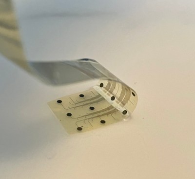
Graphene-based, high-resolution cortical brain interface (Credit: Inbrain Neuroelectronics)

Now a company called InBrain claims to have cracked making electrodes out of graphene, following a series of tests on non-human test subjects. Unlike metal probes, these carbon-based probes should be significantly more biocompatible even when used for brain stimulation as with the target goal of treating the symptoms associated with Alzheimer’s.

During the upcoming first phase human subjects would have these implants installed where they would monitor brain activity in Alzheimer’s patients, to gauge how well their medication is helping with the symptoms like tremors. Later these devices would provide deep-brain stimulation, purportedly more efficiently than similar therapies in use today. The FDA was impressed enough at least to give it the ‘breakthrough device’ designation, though it is hard to wade through the marketing hype to get a clear picture of the technology in question.

In their most recently published paper (preprint) in Nature Nanotechnology, [Calia] and colleagues describe flexible graphene depth neural probes (gDNP) which appear to be what is being talked about. These gDNP are used in the experiment to simultaneously record infraslow (<0.1 Hz) and higher frequencies, a feat which metal microelectrodes are claimed to struggle with.

Although few details are available right now, we welcome any brain microelectrode array improvements, as they are incredibly important for many types of medical therapies and research.

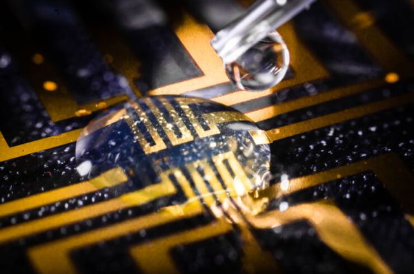
A clear droplet sits on a blue PCB with gold traces. A syringe with a drop of clear liquid sits above the droplet.

Grow Your Own Brain Electrodes

Bioelectronics has been making great strides in recent years, but interfacing rigid electrical components with biological systems that are anything but can prove tricky. Researchers at the Laboratory for Organic Electronics (LOE) have found a way to bridge the gap with conductive gels. (via Linköping University)

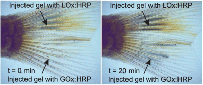
Outside the body, these gels are non-conductive, but when injected into a living animal, the combination of gel and the body’s metabolites creates a conductive electrode that can move with the tissue. This is accompanied by a nifty change in color which makes it easy for researchers to see if the electrode has formed properly.

Side-by-side images of a zebrafish tail. Both say "Injected gel with LOx:HRP" at the top with an arrow going to the upper part of the tail structure. The left says "t=0 min" and "Injected with gel GOx:HRP" along the bottom with an arrow going to the lower part of the tail structure. The tail shows darkening in the later image due to formation of bioelectrodes.

Applications for the technology include better biological sensors and enhanced capabilities for future brain-controlled interfaces. The study was done on zebrafish and medicinal leeches, so it will be awhile before you can pick up a syringe of this stuff at your local computer store, but it still offers a tantalizing glimpse of the future.

We’ve covered a few different brain electrodes here before including MIT’s 3D printed version and stentrodes.

Printed Brain Implants Give New Meaning To Neuroplasticity

3D printing has opened up a world of possibilities in plastic, food, concrete, and other materials. Now, MIT engineers have found a way to add brain implants to the list. This technology has the potential to replace electrodes used for monitoring and implants that stimulate brain tissue in order to ease the effects of epilepsy, Parkinson’s disease, and severe depression.

Existing brain implants are rigid and abrade the grey matter, which creates scar tissue over time. This new material is soft and flexible, so it hugs the wrinkles and curves. It’s a conductive polymer that’s been thickened into a viscous, printable paste.

The team took a conductive liquid polymer (water plus nanofibers of a polystyrene sulfonate) and combined it with a solvent they made for a previous project to form a conductive, printable hydrogel.

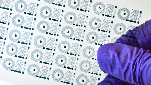
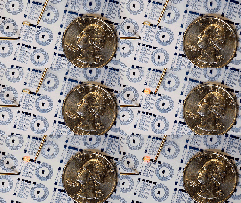
In addition to printing out a sheet of micro blinky circuits, they tested out the material by printing a flexible electrode, which they implanted into a mouse. Amazingly, the electrode was able to detect the signal coming from a single neuron. They also printed arrays of electrodes topped with little wells for holding neurons so they can study the neurons’ signals using the electrode net underneath.

This particular medical printing hack is pretty far out of reach for most of us, but not all of them are. Fire up that printer and check out this NIH-approved face shield design.