Sleep apnea is a debilitating disease that many sufferers don’t even realize they have. Those afflicted with the condition will regularly stop breathing during sleep as the muscles in their throat relax, sometimes hundreds of times a night. Breathing eventually resumes when the individual’s oxygen supply gets critically low, and the body semi-wakes to restore proper respiration. The disruption to sleep causes serious fatigue and a wide range of other deleterious health effects.
Treatment for sleep apnea has traditionally involved pressurized respiration aids, mechanical devices, or invasive surgeries. However, researchers are now attempting to develop a new drug combination that could solve the problem with pharmaceuticals alone.
For those born with certain types of congenital deafness, the cochlear implant has been a positive and enabling technology. It uses electronics to step in as a replacement for the biological ear that doesn’t quite function properly, and provides a useful, if imperfect, sense of hearing to its users.
New research has promised another potential solution for some sufferers of congenital deafness. Instead of a supportive device, a gene therapy is used to enable the biological ear to function more as it should. The result is that patients get their sense of hearing, not from a prosthetic, but from their own ears themselves.
While EEG research might help you figure out extrasensory perception, we won’t be betting on it. However, if you want to read EEG data and use an ESP32, [Cerelog-ESP-EEG] might be the right project for you. The commercial project is an 8-channel biosensing board suitable for EEG, EMG, ECG, and brain-computer interface studies. However, the company says, “We love the hacker community! We explicitly grant permission for Personal & Educational Use.” We love you too.
They do require you to agree not to sell boards you are building, and they give you schematics, but no PC board layout. That’s understandable, although we’d guess that achieving good results will require understanding how to lay out highly sensitive circuits.
As areas of uncontrolled cell growth, cancerous growth form a major problem for a multi-celled organism like us humans. Thus before they can begin to affect our long-term prospects of a continued existence, eradicating these cells-gone-wrong is essential. Unfortunately, doing so without affecting healthy cells significantly is tough. Treatments such as chemotherapy are correspondingly rough on the body, while radiation therapy is a lot more directed. Perhaps one of the more fascinating treatments involves ultrasound, with the IEEE Spectrum magazine recently covering one company providing histotripsy equipment.
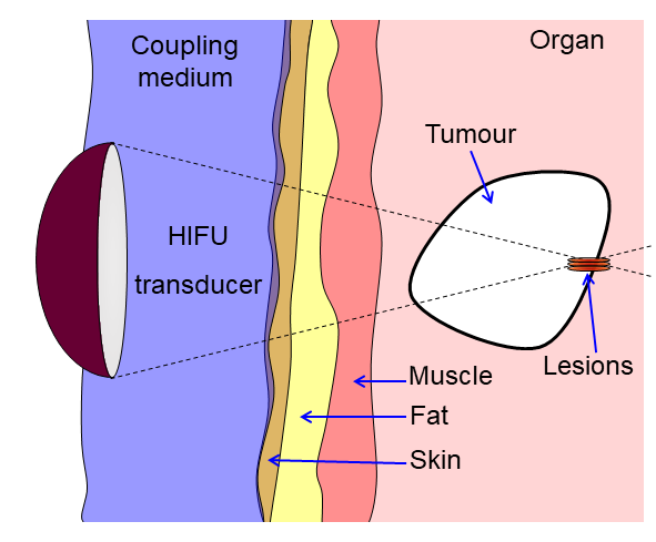
Diagram showing how HIFU can be used to destroy tissue in the body. An acoustic lens is used to focus sound to a small point in the body. (Credit: James Ross McLaughlan, Wikimedia)
Ultrasound has found many applications in the medical field far beyond imaging, with therapeutic ultrasound by itself covering a variety of methods to perform actions within the body without breaking the skin. By using high-energy ultrasound, everything from kidney stones to fat cells and cancerous cells can be accurately targeted and destroyed. For liver tumors the application of so-called histotropsy has become quite common, allowing certain types of tumors to be ablated non-invasively after which the body can handle the clean-up.
Histotropsy is a form of high-intensify focused ultrasound (HIFU) that uses either continuous or pulsed waves to achieve the desired effect, with the HIFU transducer equipped with an acoustic lens to establish a focal point. In the case of histotripsy cavitation is induced at this focal point that ends up destroying the local tissue. Beyond liver tumors the expectation is that other tumors will soon be treated in a similar manner, which could be good news for especially solid tumors.
Along with new approaches like CAR T cell immunotherapy, the prospects for cancer becoming a very treatable set of diseases would seem to be brighter than ever.
Humans have lots of basic requirements that need to be met in order to stay alive. Food is a necessary one, though it’s possible to go without for great stretches of time. Water is more important, with survival becoming difficult beyond a few days in its absence. Most of all, though, we crave oxygen. Without an air supply, death arrives in mere minutes.
The importance of oxygen is why airway management is such a key part of emergency medicine. It can be particularly challenging in cases where there is significant trauma to the head, neck, or surrounding areas. In these cases, new research suggests there may be an alternative route to oxygenating the body—through the rear.
The video covers multiple ideas on how to stabilize a hand suffering involuntary tremors. The first build involved a gyroscope, which proved unsuccessful, but led to the idea of building a reaction wheel. The concept is simple — get the reaction wheel to counteract the forces from tremors to stabilize the hand. To achieve this, an accelerometer was employed to track the movements of the arm and the hand. The magnitude of the movement was then used to control a powerful brushless motor mounted on the wrist. If the tremor was driving a hard tilt to the left, the motor would spin up to create a counter-torque, cancelling out the involuntary movement. This worked to a degree, but the resulting device was large and noisy, which made it impractical.
This thus inspired a return to earlier work involving the use of a tuned mass damper to settle tremors. The combination of some 3D printed wrist mounts along with various spring and cantilever designs… ultimately didn’t work that well. By this point, [It’s Triggy!] had noticed the tremor was mostly in the hands, while the wrist stayed steady. Thus was inspired a wrist-mounted handle for the wearer to wrap their hand around. This allowed the use of simple handheld objects like kitchen utensils, with the wearer’s own grip suppressing the tremor successfully.
Modern hospitals use a lot of computers. Architecturally speaking, they’re pretty typical machines—running the same CPUs and operating systems as any other PCs out there. However, they do tend to have some quirks when it comes to accessories and peripherals, as [tzukima] explores in a recent video.
The video starts by looking at typical power cables used with hospital computers and related equipment. In particular, [tzukima] talks about the common NEMA 5-15P to IEC-320-C13 style cable, which less sophisticated users might refer to as a kettle cord. In hospital-grade form, these cables are often constructed with translucent plug housings, with large cylindrical grips that make them easier to grip.
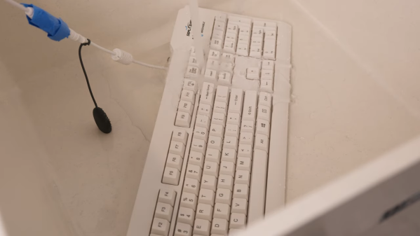
Digging further through business supply catalogs lead [tzukima] to discover further products aimed at hospital and medical users. In particular, there are a wide range of keyboards and mice that are designed for use in these environments. The most typical examples are regular peripherals that have simply been encased in silicone to make them easier to wash and disinfect where hygiene is paramount. Others, like the SealShield keyboard and mouse, use more advanced internally-sealed electronics to achieve their washable nature and IP68 ratings. These are peripherals that you can just throw in a dishwasher if you’re so inclined.