A scanning-electron micrograph is shown of a cricket's body, focusing on the head, which has been sliced off just above the eyes.

Cross-Sectioning Crickets With A Femtosecond Laser

Unlike most cutting lasers, femtosecond lasers don’t vaporize materials; rather, they produce such short, intense bursts of light that the affected region is ablated without having the chance to heat its surroundings. This makes them good at cutting away material without damaging the surroundings, something [Ben Krasnow] exploited to cut cross-sections of samples while still in a scanning-electron microscope.

In this case, the samples were crickets, and before imaging they had to be prepared. First, the bodies were soaked in glutaraldehyde to cross-link the proteins and stabilize the structure. Next, a series of solvent exchanges replaced the water in the bodies with a low-surface-tension solvent; this meant that during the next step, drying, surface tension wouldn’t distort the crickets’ internal structure. Finally, the insect bodies were charred under argon, which made the bodies conductive and more absorptive to laser light.

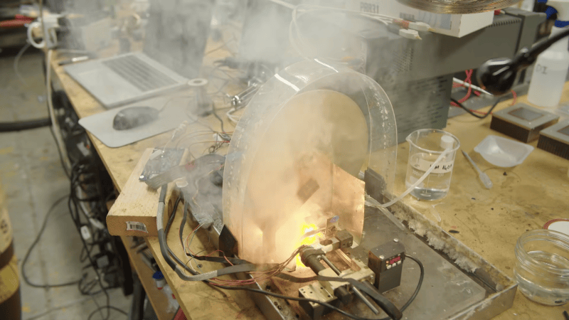
The laser itself and the scanning galvo are mounted outside the microscope, and shine in through a transparent window. To protect the detector and electron optics from a spray of ablated carbon, a servo motor swings an aluminium shutter between these and the sample while the laser is active. This caused some mysterious problems during testing: after the first ablation run, the electron microscope’s image would contain so much noise as to be unusable, but it would improve over time. As it turned out, the shutter was painted, and the other side of the paint was getting coated with charged carbon particles. This created a small capacitor which disrupted the electron optics as it discharged. Eventually, after solving this and a few other strange problems, [Ben] was able to take several time-lapse videos of the laser gradually ablating a cricket, 30 microns at a time, revealing its inner structure.

Although scanning-electron microscopes are unfortunately shard to come by, it’s still possible to restore a secondhand microscope or, as [Ben] did, build your own. Femtosecond lasers are yet more inaccessible, though they can be used to replicate themselves.

Continue reading “Cross-Sectioning Crickets With A Femtosecond Laser”

How Octopuses Hacked Their Ribosome To Become Smart

A fascinating aspect in evolutionary biology is that of convergent evolution — whereby similar structures and functions evolve independently from each other. The highly advanced nervous system of octopuses is a good example here, displaying levels of intelligence and capabilities far beyond those of other cephalopods and matching that of primates, despite no evolutionary link here. Exactly how octopuses developed this rather unique capability remained a mystery, though recent research by [Rishav Mitra] points at the rather unique ribosomes in these animals.

Ribosomes are the molecular machinery at the core of each cell that enable the synthesis of proteins. Due to their highly crucial role, they tend to remain evolutionary unchanged, which makes the big change observed in the octopus (i.e. order Octopoda) in the form of this H88 rRNA break quite remarkable.

Common octopus (<i>Octopus vulgaris</i>). (Credit: Albert Kok, Wikimedia)
Common octopus (Octopus vulgaris). (Credit: Albert Kok, Wikimedia)

This H88 break increases the accuracy of translated proteins, something that is essential for complex nervous systems as it reduces cases of misfolded proteins (proteinopathy). Because of how well-preserved ribosomes are across species, the researchers were able to run a number of experiments including a similar rRNA break in E. coli that confirmed many of the assumptions about how these octopus ribosomes performed.

Since proteinopathy results in misfolded proteins that are either useless or harmful to the organism – as seen in various human diseases – this can especially harm long-lived cells like neurons. Unsurprisingly, we can see a similar change to ribosomes in other animal groups, including that of us primates. Although the reasons for octopuses to develop more complex nervous systems wasn’t due to social pressures but rather to cope with highly complex and dynamic environments, it would seem that both types of environmental pressures led to the same convergent path, with a little ribosomal help.

Even Chemical Bonds Obey Einstein’s Relativity

Although Einstein’s Theory of Relativity is typically associated with really large and really heavy things like planets in solar systems and big things in universes in general, it turns out that even at an atomic scale its effects can be measured. These are the findings of Brown University scientists, whose measurements on very heavy elements indicate the presence of relativistic bonds.

Unfortunately the paper by [Kirk A. Peterson] et al. in Science is paywalled without a convenient ArXiv version to ogle details beyond the supplemental, but the Brown press release gives quite a few details by itself, including the use of photoelectron spectroscopy to measure the strength of the bonds between the examined nuclei.

The essential summary is that our concept of how triple bonds work may be flawed, with the assumption that there are distinct sigma and pi bonds, the latter being the awkward, weaker ‘side bonds’ where the overlapping atomic orbitals do not directly line up as with a sigma bond. As it turns out, if there’s enough mass involved, relativistic effects smudge both types of bonds together into a hybrid type of bond.

Although the sigma-pi triple bond theory still seems to hold up for lighter atomic nuclei, in the case of the examined bismuth-carbon triple bond, the typical, slightly radioactive bismuth-209 nucleus with atomic number 83 is heavy enough to affect the orbital mechanics and with it the chemical bonds that these produce.

This is an important finding, as it affects our basic understanding of how strong the bonds between certain elements are. Pi bonds are after all significantly weaker than sigma bonds, so a hybrid form would effectively make triple bonds involving a heavier element stronger than one between lighter elements.

Chromatography As Art

You may or may not remember in some ancient chemistry class studying or even performing chromatography. The short definition is using media like paper or powder to separate a mixture. It is an old technique, but [Suchir2004] is using it as an art form.

Chromatography works because the parts of the liquid mixture travel through the media at different speeds. While experimenting, [Suchir2004] noted that black ink and water perfused into constituent pigments. A butterfly ensued.

Is it art? Yes! Is it science? Well, sort of. Especially since the post does talk about how the effect works and even does some simple tests to start. This would be an excellent project for a class where some students are more motivated by art and others by science. Even with an individual kid, it might show you where their interests lie.

There’s nothing particularly difficult. A sketch pen, some paper, a coffee filter, a glue stick, and a few other household items are all you really need to get started.

Want something more practical? How about measuring caffeine content?

Newton’s Cradle Isn’t Really Perpetual

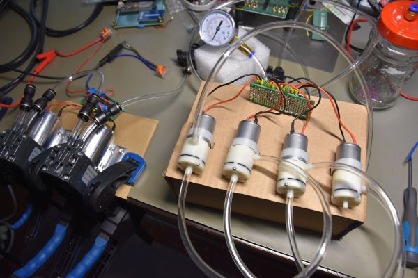
If any astute Hackaday reader saw [dongvua90]’s Newton’s cradle go on without human intervention all day long, they’d probably suspect the truth: there’s a battery and a magnet involved. But it is a nice desk piece, and you might be able to fool your less enlightened friends that you’ve discovered perpetual motion. Watch the resulting faux perpetual motion machine in action in the video below.

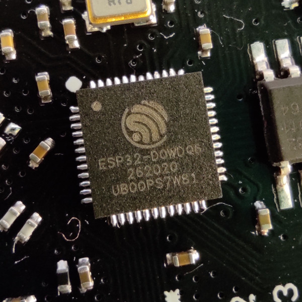
The trick is to sense the ball’s travel and inject a little electromagnetic pulse at just the right time. No problem for an ESP32 and a proximity sensor like the ones you find on some 3D printers. In fact, there’s very little custom circuitry. Everything is a module, and even the Newton’s cradle is cut out of a premade toy. A printed case and some software are really the heart of the design.

We can imagine this might be an interesting science demonstrator. Show the class the cradle with the electronics turned off, then subtly turn it on and ask the class what changed. You could even make the point by having students do it normally, while only you can get it to keep going forever, and challenge them to deduce what’s going on.

You might correctly imagine that this isn’t the first one of these we’ve seen. You can also build one that is sort of simulated.

Continue reading “Newton’s Cradle Isn’t Really Perpetual”

A series of simulations of a shape are shown, with that shape traced out in a petri dish with a laser below. The shape is roughly like a 90-degree corner bisected by a third arm.

Printing Fungal Art With Laser Control

Preservationists usually take great care to prevent fungi from appearing the world of art, but in the case of [Kexin Wang]’s Funguy project, the fungus itself is the art. It uses a laser diode to repeatedly trace an outline onto a dish of agar gel in which fungus is growing, and the photophobic fungus grows only up to the edge of the laser-traced figure, potentially creating complex designs.

This project evolved out of a research project in which they developed a computer model for fungal growth, then used its predictions and a laser to control a fungus’s growth pattern. The model has two parts: a temporal convolutional neural network which learns fungi growth patterns from a series of images, and a cellular automaton to simulate these growth patterns under different starting conditions. The cellular automaton’s rules aren’t fixed; each cell runs a small neural network which learns the rules under supervision from the convolutional network. By training these networks on images of the growth stages of three different fungi, it was able to realistically predict the different growth patterns of the different species.

To actually control the growth pattern, the researchers tried a series of different wavelengths and laser powers; shorter wavelengths tended to work better, with a 405 nm laser working best. The growth model complemented the laser setup by predicting in which areas the growth medium had run out of nutrients. Since fungus would no longer spread in these regions, the laser no longer needed to trace these sections. The Funguy kit’s laser system itself is similar to a laser engraver, with an XY-kinematic system seemingly built from a DVD drive frame. It uses fungi from the Mucor genus, though it can print with other photophobic microorganisms, such as slime molds.

This project seems aimed at artistic and educational uses, but considering the various electronic parts that have been made of fungi, more functional applications should be possible.

Mechanosynthesis Of Atomic Carbon Structures Using Inverted-Mode STM

Generally chemical synthesis involves putting a variety of compounds together in an environment where they will react and self-assemble into the desired product. You could also imagine simply putting the atoms in the right place: direct mechanical manipulation. This mechanosynthesis is however not that simple, despite the deceptive appearance of those ball-and-stick representations in high school chemistry class.

This is demonstrated in a recent (pre-publication) study by [Megan Cowie] et al. using inverted-mode STM. Using a scanning tunneling microscope (STM) you can measure a surface on a nanoscale, with the inversed principle used in inverted-mode STM (IM-STM) to physically move individual molecules. In the paper the construction of carbon-based 3D structures using IM-STM is demonstrated.

In the paper it is demonstrated how C2 units can be moved using the tip of an IM-STM setup for subsequent polyyne structure construction through C-C bond formation at the target site. Although it’s not quite yet the leap into Neal Stephenson’s The Diamond Age with its science-based matter compilers – i.e. molecular assemblers – it’s definitely another step closer to making advanced feats of nanotechnology a part of every day life.