Monitor For Bedridden Patients Aims To Improve Care

One of the joys of being a Maker and Hacker is solving problems and filling needs. When you can do both, well, that’s something special. [rodrigo.mejiasz]’s project surely fits into that special category of solving a problem and filling a dire need with his Bedridden Patient Monitor.

While [Rodrigo]’s project page does not specify his motivation for creating this project, one only needs to look as far as their local hospital ward or senior care facility to understand why this device is so wonderful. Healthcare workers and caregivers are stretched paper thin, and their attention is being constantly interrupted.

This is where the Bedridden Patient Monitor comes in. A healthy person can reposition themselves if they are uncomfortable, but bedridden patients cannot. It’s not just that a bedridden patient is unable to get out of bed, but that they are unable to move themselves without assistance. The result is a great amount of pain. And if left unchecked, pressure sores can be the result. These are not only extremely unpleasant, but an added danger to a patients health.

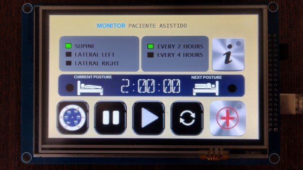
The Bedridden Patient Monitor steps in and provides not just an egg-timer like alert, but helps caregivers track a patients position in bed across even several working shifts. This ensures a continuity of care that might otherwise be easy to miss.

The beauty of this build is in its application but also its simplicity: it’s just an Arduino Mega, a TFT shield with its Micro SD card, and the touch screen itself. A few LED’s and a buzzer take care of alerts. A thoughtfully configured interface makes the devices use obvious so that staff can make immediate use of the monitor.

Makers have a long history diving into the medical field, such as this stab wound treatment device that won the Dyson award in 2021.

Continue reading “Monitor For Bedridden Patients Aims To Improve Care”

3D Printing A Real Heart

As 3D printing becomes more and more used in a wide range of fields, medical science is not left behind. From the more standard uses such as printing medical equipment and prosthetics to more advanced uses like printing cartilages and bones, the success of 3D printing technologies in the medical field is rapidly growing.

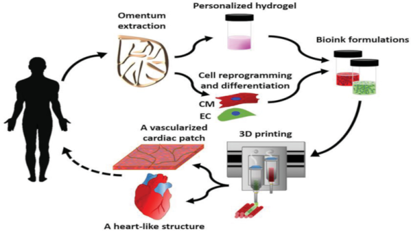
One of the last breakthrough is the world’s first 3D vascularised engineered heart using the patient’s own cells and biological materials. Until now, scientists have only been successful in printing only simple tissues without blood vessels. Researchers from Tel Aviv University used the fatty tissue from patients to separate the cellular and acellular materials and reprogrammed the cells become pluripotent stem cells. The extracellular matrix (ECM) was processed into a personalized hydrogel that served as the basis from the print.

This heart is made from human cells and patient-specific biological materials. In our process these materials serve as the bioinks, substances made of sugars and proteins that can be used for 3D printing of complex tissue models… At this stage, our 3D heart is small, the size of a rabbit’s heart, but larger human hearts require the same technology.

After being mixed with the hydrogel, the cells were efficiently differentiated to cardiac or endothelial cells to create patient-specific, immune-compatible cardiac patches with blood vessels and, subsequently, an entire heart that completely matches the immunological, cellular, biochemical and anatomical properties of the patient. The difficulty of printing full-blown organs were being tackled for a long time and we already talked about it in the past.

The development of this technology may completely solve both the problem of organ compatibility and organ rejection.


Towards Low Cost Biomedical Imaging

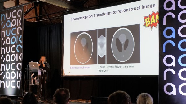
Medical imaging is one of the very best applications of technology — it allows us to peer inside of the human body without actually performing surgery. It’s non-destructive testing to the extreme, and one of the more interesting projects we’ve seen over the past year uses AC currents and an infinite grid of resistors to image the inside of a living organism. It’s called Spectra and it is the brainchild of [Jean Rintoul]. Her talk at the Hackaday Superconference is all about low cost and open source biomedical imaging.

We’ve seen some interesting medical imaging hacks in the Hackaday Prize over the years. There have been vein finders and even a CT scanner, but when it comes to biomedical imaging, the Spectra project is something different. Right now, it’s just good enough to image organs while they’re still inside your body, and there’s still a lot of potential to do more. Let’s take a closer look a how this works.

Continue reading “Towards Low Cost Biomedical Imaging”

Infection? Your Smartphone Will See You Now

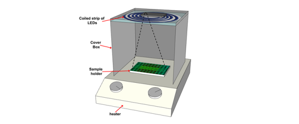
When Mr. Spock beams down to a planet, he’s carrying a tricorder, a communicator, and a phaser. We just have our cell phones. The University of California Santa Barbara published a paper showing how an inexpensive kit can allow your cell phone to identify pathogens in about an hour. That’s quite a feat compared to the 18-28 hours required by traditional methods. The kit can be produced for under $100, according to the University.

Identifying bacteria type is crucial to prescribing the right antibiotic, although your family doctor probably just guesses because of the amount of time it takes to get an identification through a culture. The system works by taking some — ahem — body fluid and breaking it down using some simple chemicals. Another batch of chemicals known as a LAMP reaction mixture multiplies DNA and will cause fluorescence in the case of a positive result.

Continue reading “Infection? Your Smartphone Will See You Now”

MRI To 3D Print Gets Much Faster

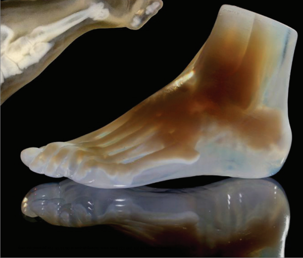
A surprising use of 3D printing has been in creating life-like models of human body parts using MRI or CT scans. Surgeons and other medical professionals can use models to plan procedures or assist in research. However, there has been a problem. The body is a messy complex thing and there is a lot of data that comes out of a typical scan. Historically, someone had to manually identify structures on each slice — a very time-consuming process — or set a threshold value and hope for the best. A recent paper by a number of researchers around the globe shows how dithering scans can vastly improve results while also allowing for much faster processing times.

As an example, a traditional workflow to create a 3D printed foot model from scan data took over 30 hours to complete including a great deal of manual intervention. The new method produced a great model in less than an hour.

Continue reading “MRI To 3D Print Gets Much Faster”

Mae Jemison And The Final Frontier

From the time Mae Jemison was a little girl, she was convinced that she would go to space. No one could tell her otherwise. She was sure that space travel would be as common as air travel by the time she was an adult. That prediction didn’t pan out, but that confidence combined with her intellect, curiosity, and the above-average encouragement of her parents drove Mae to do everything she wanted, including space travel.

Some people might become a doctor or a researcher, a dancer or an astronaut. But Mae became all of these things. Not everyone supported her non-traditional path—many people just pick a career and stick with it. Her path is impressive and through it all she gained a really interesting perspective on how education is approached, and what effects that approach has on society. After practicing medicine, joining a shuttle mission, appearing in Star Trek, and retiring from NASA, she became a voice for minority students and an advocate for integrating the arts and sciences in the standard curriculum.

Continue reading “Mae Jemison And The Final Frontier”

Hackaday Prize Entry: Stroke Rehabilitation Through Biofeedback

Students at Purdue University’s Weldon School of Biomedical Engineering created ExoMIND, an Arduino-powered glove that helps a stroke victim recover by tracking the range of motion the patient experiences.

A set of 7 accelerometers in the fingers, wrist, and forearm track the range of movements the patient is experiencing with that hand. An accelerometer on the back of the hand serving as a reference. Meanwhile, an EMG sensor working with a conductive fabric sleeve to measure muscle activity. The user follows a series of instructions dished out by an interactive software program, allowing the system to test out the patient’s range of motion at the beginning of the regime as well as to record whether any improvement was noted at the end. The data is used by a physical therapist to personalize the treatment plan. The interactive program also raises the possibility of patients self-directing their exercises with the ExoMIND telling them how to adjust their motion to get the most out of the experience.

Produced as part of the university’s MIND Biomedical Engineering Club, the ExoMIND prototype was designed by three interdisciplinary teams focusing on electronics, materials, and programming, respectively.

Continue reading “Hackaday Prize Entry: Stroke Rehabilitation Through Biofeedback”