Homemade Raman Laser Is Shaken, Not Stirred

You wouldn’t think that shaking something in just the right way would be the recipe for creating laser light, but as [Les Wright] explains in his new video, that’s pretty much how his DIY Raman laser works.

Of course, “shaking” is probably a gross oversimplification of Raman scattering, which lies at the heart of this laser. [Les] spends the first half of the video explaining Raman scattering and stimulated Raman scattering. It’s an excellent treatment of the subject matter, but at the end of the day, when certain crystals and liquids are pumped with a high-intensity laser they’ll emit coherent, monochromatic light at a lower frequency than the pumping laser. By carefully selecting the gain medium and the pumping laser wavelength, Raman lasers can emit almost any wavelength.

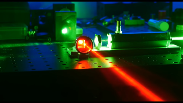
Most gain media for Raman lasers are somewhat exotic, but luckily some easily available materials will work just fine too. [Les] chose the common solvent dimethylsulfoxide (DMSO) for his laser, which was made from a length of aluminum hex stock. Bored out, capped with quartz windows, and fitted with a port to fill it with DMSO, the laser — or more correctly, a resonator — is placed in the path of [Les]’ high-power tattoo removal laser. Laser light at 532 nm from the pumping laser passes through a focusing lens into the DMSO where the stimulated Raman scattering takes place, and 628 nm light comes out. [Les] measured the wavelengths with his Raspberry Pi spectrometer, and found that the emitted wavelength was exactly as predicted by the Raman spectrum of DMSO.

It’s always a treat to see one of [Les]’ videos pop up in our feed; he’s got the coolest toys, and he not only knows what to do with them, but how to explain what’s going on with the physics. It’s a rare treat to watch a video and come away feeling smarter than when you started.

Continue reading “Homemade Raman Laser Is Shaken, Not Stirred”

SHERLOC And The Search For Life On Mars

Humanity has been wondering about whether life exists beyond our little backwater planet for so long that we’ve developed a kind of cultural bias as to how the answer to this central question will be revealed. Most of us probably imagine that NASA or some other space agency will schedule a press conference, an assembled panel of scientific luminaries will announce the findings, and newspapers around the world will blare “WE ARE NOT ALONE!” headlines. We’ve all seen that movie before, so that’s the way it has to be, right?

Probably not. Short of an improbable event like an alien spacecraft landing while a Google Street View car was driving by or receiving an unambiguously intelligent radio message from the stars, the conclusion that life exists now or once did outside our particular gravity well is likely to be reached in a piecewise process, an accretion of evidence built up over a long time until on balance, the only reasonable conclusion is that we are not alone. And that’s exactly what the announcement at the end of last year that the Mars rover Perseverance had discovered evidence of organic molecules in the rocks of Jezero crater was — another piece of the puzzle, and another step toward answering the fundamental question of the uniqueness of life.

Discovering organic molecules on Mars is far from proof that life once existed there. But it’s a step on the way, as well as a great excuse to look into the scientific principles and engineering of the instruments that made this discovery possible — the whimsically named SHERLOC and WATSON.

Continue reading “SHERLOC And The Search For Life On Mars”

See Cells In A New Light With A DIY Fluorescence Microscope

Ever since a Dutch businessman peered into the microscopic world through his brass and glass contraption in the 1600s, microscopy has had a long, rich history of DIY innovation. This DIY fluorescence microscope is another step along that DIY path that might just open up a powerful imaging technique to amateur scientists and biohackers.

In fluorescence microscopy, cells are treated with various fluorescent dyes that can be excited with light at one wavelength and emit light at another. But as [Jonathan Bumstead] points out, fluorescence microscopes are generally priced out of the range of biohackers. His homebrew scope levels the playing field a bit. The trick is to use 3D-printed parts to kit out commonly available digital cameras – a USB microscope, a DSLR, or even a smartphone camera. Excitation is provided by a ring of Nexopixel LEDs, while a movable rack holds a filter that blocks the excitation wavelength but allows the emission wavelength to pass through to the camera. He demonstrates the technique by staining some threads with fluorescent ink from a highlighter marker and placing them on a sheet of tissue paper; in conventional bright-field mode, the threads all but disappear into the background, but jump right out under fluorescence.

Ith’s true that the optics are not exactly lab quality, and the microscope is currently only set up to do reflectance imaging as opposed to the more typical transmissive mode where the light passes through the sample. That’s an easy fix, though, and reflectance mode is still useful. We’ve seen fluorescence microscopy get quite complex before, but this simple scope might be enough to get a biohacker started.

Continue reading “See Cells In A New Light With A DIY Fluorescence Microscope”