When you think of Florence Nightingale, you probably imagine a nurse with a lamp, comforting soldiers. Indeed, Florence is considered the mother of modern nursing. But she also made serious contributions in statistical data analysis, and used the diagram named after her, the Nightingale rose diagram, to convince the British Parliament to enact sanitation reforms that saved hundreds of thousands, if not millions, of lives.
During the Crimean war, Florence worked around the clock as head nurse in an overcrowded field hospital. But she also found time to create graphs to illustrate the terrible conditions of that field hospital to members of British Parliament. The sanitation reforms she led greatly improved the life of the soldiers in battle, and widespread adoption of her hygienic practices vastly reduced mortality rates of humanity in general.
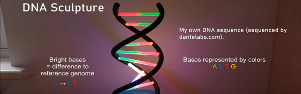
It has recently been possible to pay a service a little bit of money and learn more about your own DNA. You might find out you really aren’t Italian after all or that you are more or less susceptible to some ailments. [Paul Klinger] had his DNA mapped and decided to make a sculpture representing his unique genetic code. The pictures are good, but the video below is even better.
The project requires a DNA sequencing, a 3D printer, and a Raspberry Pi Zero. Oh, you can probably guess you need a lot of RGB LEDs, too. Of course, the display doesn’t show the whole thing at one time — your DNA pattern scrolls across the double helix.
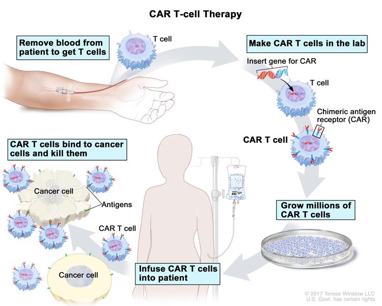
One of the human body’s greatest features is its natural antivirus protection. If your immune system is working normally, it produces legions of T-cells that go around looking for abnormalities like cancer cells just to gang up and destroy them. They do this by grabbing on to little protein fragments called antigens that live on the surface of the bad cells and tattle on their whereabouts to the immune system. Once the T-cells have a stranglehold on these antigens, they can release toxins that destroy the bad cell, while minimizing collateral damage to healthy cells.
This rather neat human trick doesn’t always work, however. Cancer cells sometimes mask themselves as healthy cells, or they otherwise thwart T-cell attacks by growing so many antigens on their surface that the T-cells have no place to grab onto.
Medical science has come up with a fairly new method of outfoxing these crafty cancer cells called CAR T-cell therapy. Basically, they withdraw blood from the patient, extract the T-cells, and replace the blood. The T-cells are sent off to a CRISPR lab, where they get injected with a modified, inactive virus that introduces a new gene which causes the T-cells to sprout a little hook on their surface.
This hook, which they’ve dubbed the chimeric antigen receptor (CAR), allows the T-cell to chemically see through the cancer cells’ various disguises and attack them. The lab multiplies these super soldiers and sends them back to the treatment facility, where they are injected into the patient’s front lines.
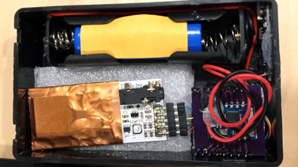
Those of us who have not been in that position can only imagine the anguish of learning that your teenager has cancer. This happened to [Rob], whose child was diagnosed with papillary thyroid cancer. It’s a condition that can be treated with surgery followed by a course of radioactive iodine to kill any remaining cancer cells. During iodine treatment, the patient is radioactive enough that other people must maintain a distance of 3m from them, and as a learning exercise for both father and teen he created and refined the design of a portable wireless radioactivity monitor.
There are a variety of sensors for radiation monitoring including the well-known Geiger–Müller tube, but he settled on a PIN photodiode based sensor supplied by radiation-watch.org. This sensor is not at its most sensitive at the energy levels emitted by the iodine isotope used in the treatment, but the relatively high intensity of the radiation meant that enough would register for a useful reading to be taken. The sensor board he was mated to an ESP8266 module. [Rob] went through three iterations of the balance of the hardware before settling on a lithium-ion battery and a plastic case.
On the software side, the ESP connects to an MQTT server, from which a CSV file of data is derived. On a computer, the CSV data is collected and plotted to a graph. The data take during treatment clearly shows the reduction in radiation following the isotope’s half-life. The graph isn’t perfect though, there is a gap due to the second prototype’s batteries running flat
From his epilogue it appears that his son has recovered, and we wish them further good health. The details have been published in the hope that other young people facing the same trial might benefit from building their own radiation monitor.
Welcome to the first installment of Inputs of Interest. In this column, we’re going to take a look at various input devices and methods, discuss their merits, give their downsides a rundown, and pontificate about the possibilities they present for hackers. I’ll leave it open to the possibility of spotlighting one particular device (because I already have one in mind), but most often the column will focus on input concepts.
Some inputs are built for having fun. Some are ultra-specific shortcuts designed to do work. Others are assistive devices for people with low mobility. And many inputs blur the lines between these three ideas. This time on Inputs of Interest, we’re going to chew on the idea of oral inputs — those driven by the user’s tongue, teeth, or both.
Unless you’ve recently bitten it, burned it, or had it pierced, you probably don’t think much about your tongue. But the tongue is a strong, multi-muscled organ that rarely gets tired. It’s connected to the brain by a cranial nerve, and usually remains undamaged in people who are paralyzed from the neck down. This makes it a viable input-driving option for almost everyone, regardless of ability. And yet, tongues and mouths in general seem to be under-utilized as input appendages.
Ideally, any input device should be affordable and/or open source, regardless of the driving appendage. Whether the user is otherwise able-bodied or isn’t, there’s no reason the device shouldn’t be as useful and beautiful as possible.
Assistive technology is extremely fertile ground for hackers to make a difference, because of the unique requirements of each user and the high costs of commercial solutions. [Nick] has been working on Earswitch, an innovative assistive tech switch that can be actuated using voluntary movement of the middle ear muscle.
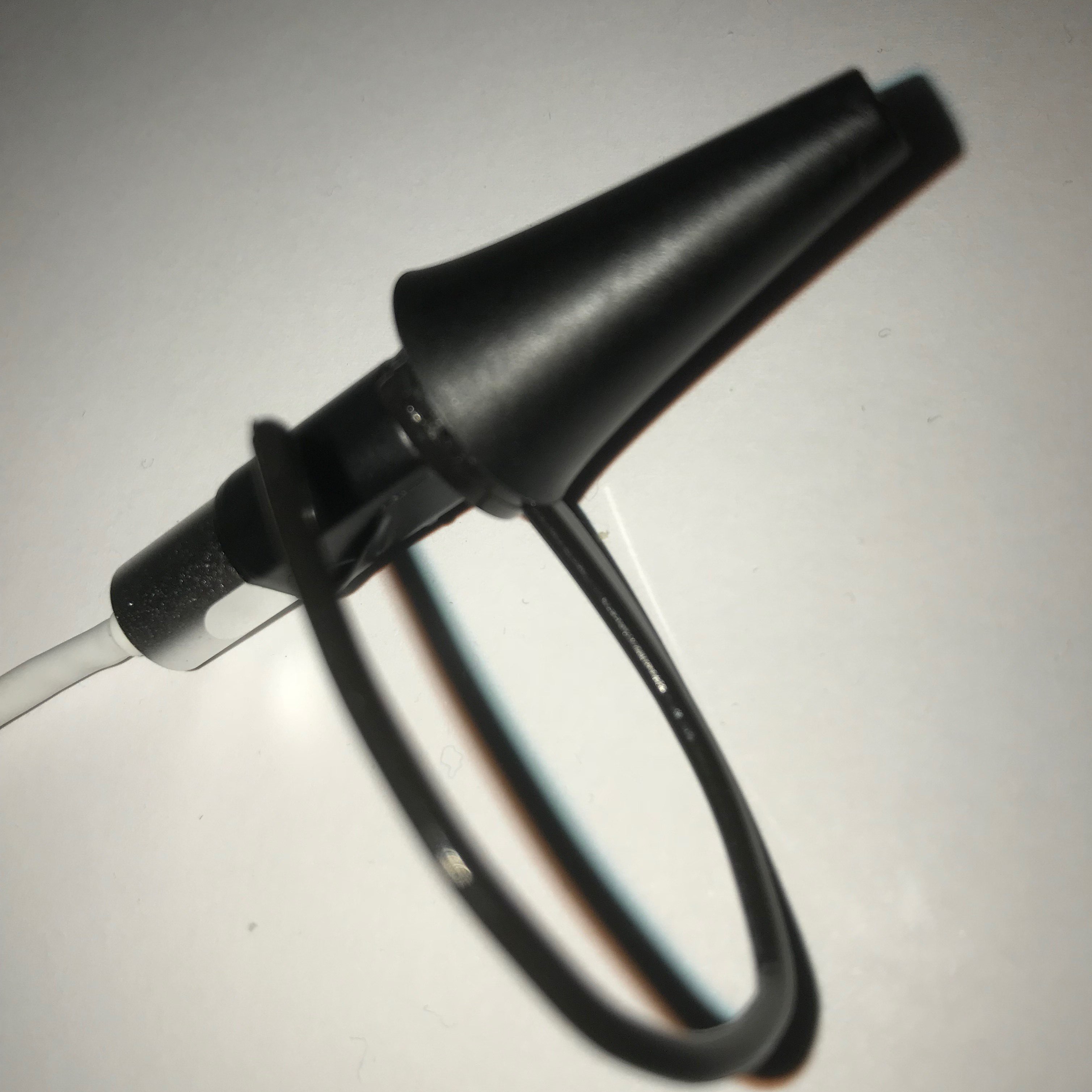
Most people don’t know they can contract their middle ear muscle, technically called the tensor tympani, but will recognise it as a rumbling sound or muffling effect of your hearing when yawning or tightly closing eyes. Its function is actually to protect your hearing from loud sounds screaming or chewing. [Nick] ran a survey and found that 75% can consciously contract the tensor tympani and 17% of can do it in isolation from other movements. Using a cheap USB auroscope (an ear camera like the one [Jenny] reviewed in November), he was able to detect the movement using iSpy, an open source software package meant for video surveillance. The output from iSpy is used to control Grid3, a commercial assistive technology software package. [Nick] also envisions the technology being used as a control interface for consumer electronics via earphones.
With the proof of concept done, [Nick] is looking at ways to make the tech more practical to actually use, possibly with a CMOS camera module inside a standard noise canceling headphones. Simpler optical sensors like reflectance or time-of-flight are also options being investigated. If you have suggestions for or possible use case, drop by on the project page.
Assistive tech always makes for interesting hacks. We recently saw a robotic arm that helps people feed themselves, and the 2017 Hackaday Prize has an entire stage that was focused on assistive technology.
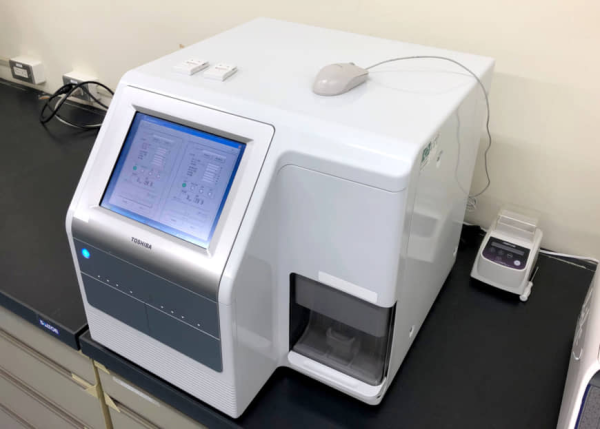
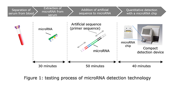
Can you imagine a near future where your family doctor can effectively prick your finger and test you for a dozen or so types of cancer? Currently, cancer detection is a time-consuming and expensive process. Existing methods of screening for cancer usually involve taking a whole lot of blood and running tests that cost thousands of dollars. But Toshiba has created a cancer-detecting machine that sounds like something straight out of science fiction.
The machine is about the size of a small office copier, and it looks like one, too. But this small machine can do some powerful tricks. Toshiba claims that the machine can detect 13 types of cancer from a single drop of blood with 99% accuracy. What’s more impressive is that it can do this under two hours, as opposed to days or weeks depending on laboratory backlog. Most importantly, they are aiming to do this entire battery of tests for about $180. Ideally, this machine will do everything that current blood cancer detection equipment does, just better, faster, and with fewer resources.
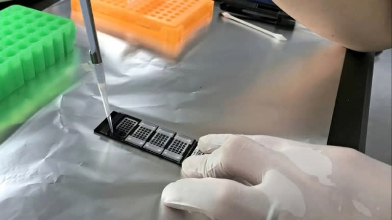
Some of the cancers the machine can test for have been previously difficult to detect, like ovarian, pancreatic, and esophageal cancer. But this machine can screen for all three of these — great news for early detection of these ravaging cancers — as well as breast, prostate, gastric, colon, liver, biliary tract, bladder, lung, brain, and sarcoma. The only catch is that the machine can’t pinpoint which cancer exactly, it only knows if microRNA one or more of the 13 came up.
So, how does it work? Cancer cells secrete certain types of microRNA into the bloodstream that healthy cells don’t. The machine works by assessing the different types of microRNA that show up in the sample drop, and studying their concentrations. Their work builds on that of Toray Industries, who announced earlier this year that they had made a cancer-detection chip based on microRNA accumulation that is 95% accurate. Development of this chip follows on the heels of research that finds testing for microRNA in bloodwork has the potential to detect cancers in earlier stages, and in some cases like for bowel cancer, with a much less invasive testing procedure.
Toshiba, in partnership with the National Cancer Center Research Institute and Tokyo Medical University will conduct a trial of the machine next year. If the trial is successful, they hope to commercialize it soon after.