ARPA-H Moonshot Project Aims To Enable 3D Printing Of Human Organs

The field of therapeutic cloning has long sought to provide a way to create replacement organs and tissues from a patient’s own cells, with the most recent boost coming from the US Advanced Research Projects Agency for Health (ARPA-H) and a large federal contract awarded to Stanford University.
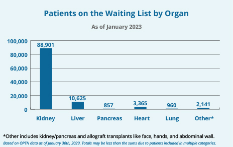
Patients on the organ donation waiting list in the US (Source: HRSA)

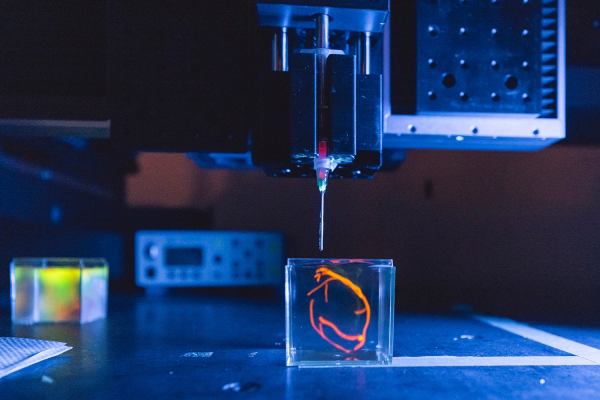
The creatively named Health Enabling Advancements through Regenerative Tissue Printing (HEART) project entails a 26.3 million USD grant that will be used to create a functioning bioprinter backed by a bank of bioreactors. Each bioreactor will cultivate a specific type of cell, which will then be ‘printed’ in its proper place to gradually build up the target organ or tissue. The project’s five year goal is the printing of a fully functioning human heart and implanting it into a pig.

Assuming this is successful, the general procedure can then be refined to allow for testing with human patients, as well as the bioprinting of not just hearts, but also lungs, kidneys and much more. The lead investigator at Stanford University, [Mark Skylar-Scott], cautions that use with human patients is likely to be still decades off. But the lifesaving potential of this technology, once matured, is staggering. This is highlighted by data from the US HRSA, with over 42,000 transplants in 2022 in the US alone, with over a hundred-thousand patients waiting and 17 people who die each day before an organ becomes available.
Bioadhesive Polymer Semiconductors For In-Vivo Sensors

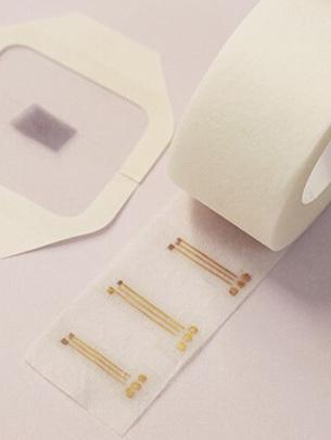
The bioadhesive electrodes on a roll.

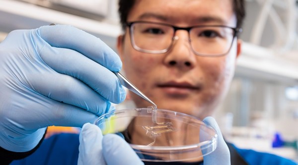
What do you do when you want to stick an electrode or even an couple of sensors to an internal organ, such as a heart? Generally you’d use some kind of special adhesive, or sutures to ensure that the item remains firmly in place and doesn’t migrate to somewhere else within the chest cavity or among the intestines. According to a new study (press release) by Nan Li and colleagues in Science there may however be a more elegant method, using bioadhesive polymers.

The double-network copolymer is designed so that once put in the desired location it soaks up moisture and provides a dry interface for its bioadhesive properties. In addition, the resulting material is electrically conductive, with a measured charge-carrier mobility of ~1 square centimeter per volt per second.

Using thus manufactured electrodes were applied to both an isolated rat heart and in vivo rat muscles to measure electrical currents produced by each respective tissue type. The authors of the study envision that using this technology more complicated interfaces and sensors can be developed that would interface directly with organs and related. The claimed biocompatibility would also allow for such devices to be left in-situ for extended periods of time, which could be a boon for a wide range of medical conditions where continuous monitoring is a crucial element.

Metamaterial Enables Topological Pumping Of Elastic Surface Waves

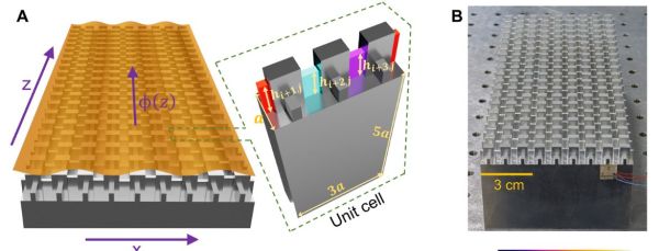
Although it is generally assumed that surface elastic waves (vibrations) — such as those of earthquakes — will travel mostly unimpeded until their energy dissipates, there are ways to ‘steer’ this energy using metamaterials.

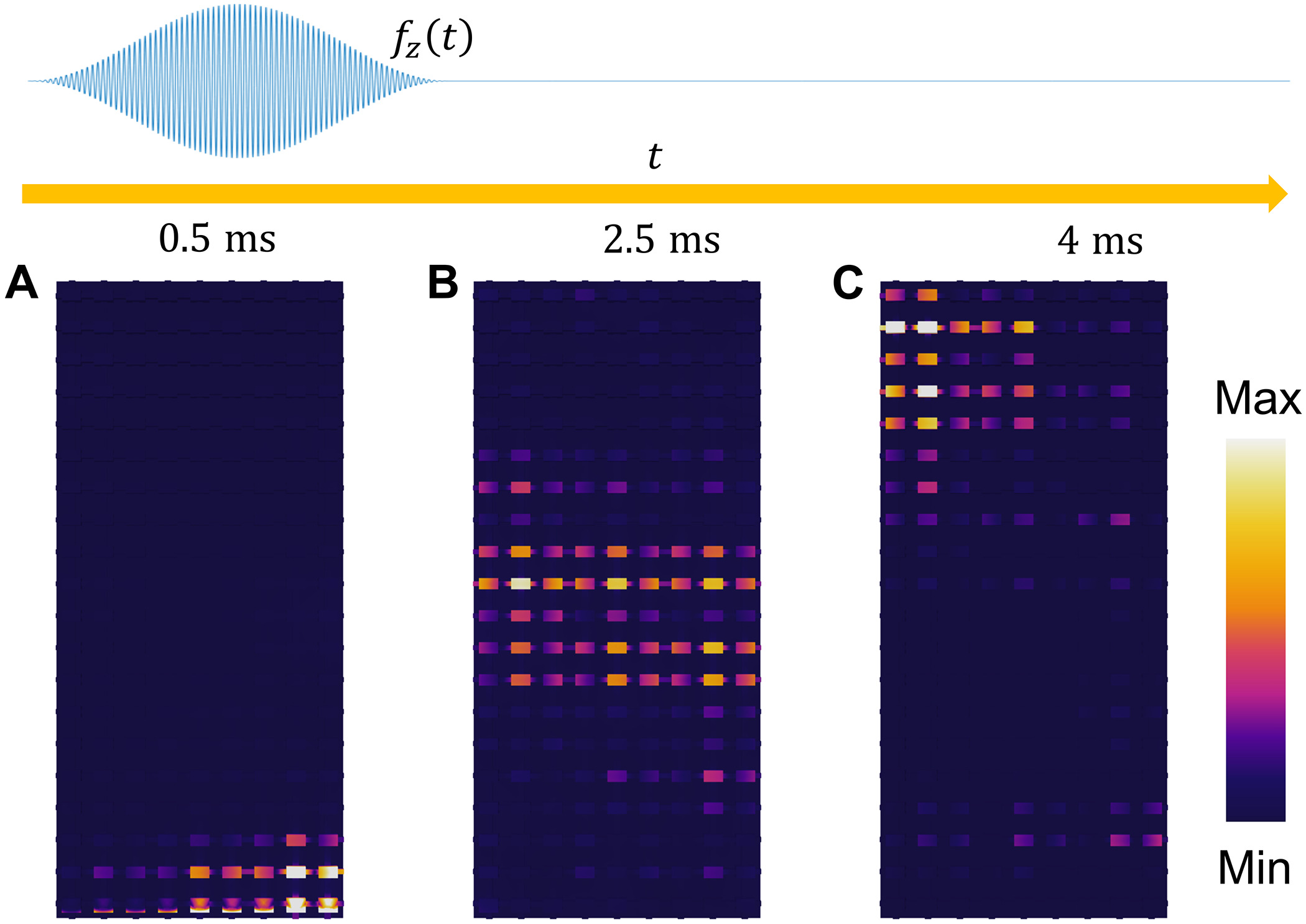
Time response of the topological surface wave transport.(A to C). The magnitude of total displacement field at 0.5 ms, 2.5 ms, and 4 ms, respectively. A 50-cycle tone burst signal centered at 41.88 kHz is simulated on the bottom supercell. (Wang et al., 2023)
Time response of the topological surface wave transport.
(A to C). The magnitude of total displacement field at 0.5 ms, 2.5 ms, and 4 ms, respectively. A 50-cycle tone burst signal centered at 41.88 kHz is simulated on the bottom supercell. (Wang et al., 2023)

A recent study by [Shaoyun Wang] and colleagues in Science Advances details how a carefully modelled grouping of columns creates what is termed a synthetic dimension. In their experimental setup, it is demonstrated how an applied wave is guided across the metamaterial, rather than spreading out the way which we would expect to see in conventional materials.

Interestingly, in the paper it is also demonstrated how the same technique can be used to create a wave-splitter that diverts the wave energy in two distinct directions. Due to the innate resistance of this type of structure to defects, manufacturing it is not too complicated.

In this experiment the metamaterials were milled out of a block of aluminium on a CNC mill, which makes it seem eminently realistic that it could be scaled up and translated to other applications. Conceivably annoyances like vibrations from road traffic and heavy machinery, all the way up to the destructive energies of earthquakes could one day be reduced, redirected or even extinguished using structures as demonstrated here.

Femtosecond Laser Clones Itself In Glass

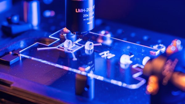
When researchers at the Galatea laboratory in Switzerland set out to create a femtosecond laser in glass they weren’t certain it was going to work. To be precise, their goal was to create a femtosecond laser cavity using carefully aligned optics. Rather than using the traditional, discrete method, they used a commercial femtosecond laser to carve out the elements of the optical cavity in glass. The choice for glass came down to the low thermal expansion of this material, and it being transparent for the optical frequencies being targeted.

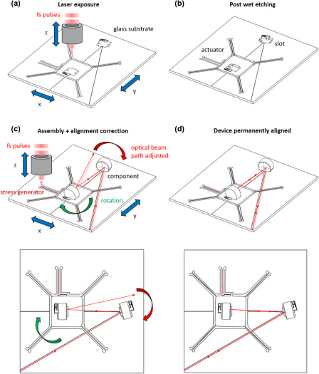
Generic concept of an “all-glass” optical device, with the various stages of fabrication. (Credit: Antoine Delgoffe et al., 2023)

Even after using the existing laser to create the rough laser cavity, the resulting optical mirrors were not aligned properly, but this was all part of the plan.

By also adding slots that created a flexure mechanism, brief laser pulses could be used to gradually adjust the mirrors to create the perfect alignment. During subsequent testing of the newly created laser cavity it was found to be operating as expected. The original femtosecond laser had successfully created a new femtosecond laser.

Perhaps the most tantalizing aspect of this research is that this could enable much faster and ultimately cheaper production of such laser systems, especially once the tedious and currently completely manual mirror alignment procedure is automated. In addition, it raises the prospect of producing other types of optics including splitters and guides in a similar manner.

The Path To Profiling Extraterrestrial Atmospheres With Astrophotonics

A major part of finding extraterrestrial life is to be able to profile the atmosphere of any planets outside of our solar system. This is not an easy task, as these planets are usually found through the slight darkening of their star as they pass in front of it (transition). Although spectroscopy is the ideal way to profile the chemical composure of such a planet, having a massive, extremely bright star right next to the planet is more than enough to completely overpower the faint light reflecting off the planet’s surface and through its atmosphere. This is a major issue that the upcoming Habitable Exoplanet Imaging Mission (HabEx, also called the Habitable Worlds Observatory, or HWO) hopes to address using a range of technologies, including a coronagraph that should block out most of the stellar glare.

While this solves much of the issue, there are still a range of issues which the new field of astrophotonics seeks to address, as detailed in a recent paper by Nemanja Jovanovic and colleagues. This involves not only profiling chemical compositions, but also increasing the precision when monitoring for planet transit events using e.g. semiconductors-based laser frequency combs. These are generally combined with a spectral flattener, which in experimental on-chip form are significantly less bulky than previous setups, to the point where they don’t necessarily have to be Earth-based.

Continue reading “The Path To Profiling Extraterrestrial Atmospheres With Astrophotonics” →

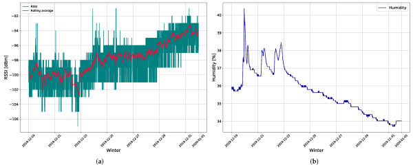
The measurement results of: (a) RSSI in dBm collected from gateway 2 and (b) soil moisture during the winter period. (Credit: Maja Škiljo et al., 2022)

Using LoRa Nodes As Soil Moisture Sensing Antennas

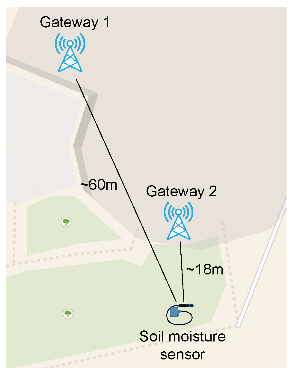
Implementation of LoRaWAN-based soil moisture sensing device. (Credit: Maja Škiljo et al., 2022)

Although we generally think of Internet of Things (IoT) and similar devices as things that are scattered around above ground, there are plenty of reasons to also have such devices underground. These so-called IoUT devices are extremely useful when it comes to monitoring underground structures, but communication via radiowaves is obviously impacted when soil is in the way. Although there are ways to get around this, a 2022 paper by Maja Škiljo and colleagues in Sensors covers an interesting way to make use of this signal attenuation property of changing moisture levels in soil.

By quantifying the exact attenuation of the signal received at the gateways, they were able to determine the soil moisture levels around the LoRa node which had been buried at a depth of approximately 14 centimeters. This LoRa node used off-the-shelf components consisting of an ATmega328P-based Arduino Pro Mini and SX1276-based RFM95W LoRa module with a spring antenna.

During experimentation in- and outdoors it was determined that a narrowband, printed (PCB) antenna was optimal for soil moisture sensing purposes. Other than the interesting question of how to keep soil moisture sensing nodes like this powered up over long periods of time (perhaps periodic retrieval to replenish the battery), this would seem to be a very interesting way to monitor the soil moisture levels in something like a field, where each node can provide its own ID and the received signal providing the relevant data in the form of the SNR and other parameters recorded by the gateway.

(Heading image: The measurement results of: (a) RSSI in dBm collected from gateway 2 and (b) soil moisture during the winter period. (Credit: Maja Škiljo et al., 2022) )

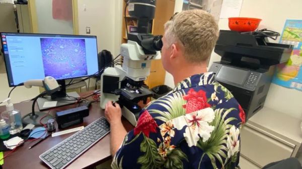
Dr. Niels Olson uses the Augmented Reality Microscope. (Credit: US Department of Defense)

Google’s Augmented Reality Microscope Might Help Diagnose Cancer

Despite recent advances in diagnosing cancer, many cases are still diagnosed using biopsies and analyzing thin slices of tissue underneath a microscope. Properly analyzing these tissue sample slides requires highly experienced and skilled pathologists, and remains subject to some level of bias. In 2018 Google announced a convolutional neural network (CNN) based system which they call the Augmented Reality Microscope (ARM), which would use deep learning and augmented reality (AR) to assist a pathologist with the diagnosis of a tissue sample. A 2022 study in the Journal of Pathology Informatics by David Jin and colleagues (CNBC article) details how well this system performs in ongoing tests.

For this particular study, the LYmph Node Assistant (LYNA) model was investigated, which as the name suggests targets detecting cancer metastases within lymph node biopsies. The basic ARM setup is described on the Google Health GitHub page, which contains all of the required software, except for the models which are available on request. The ARM system is fitted around an existing medical-grade microscope, with a camera feeding the CNN model with the input data, and any relevant outputs from the model are overlaid on the image that the pathologist is observing (the AR part).

Although the study authors noted that they saw potential in the technology, as with most CNN-based systems a lot depends on how well the training data set was annotated. When a grouping of tissue including cancerous growth was marked too broadly, this could cause the model to draw an improper conclusion. This makes a lot of sense when one considers that this system essentially plays ‘cat or bread’, except with cancer.

These gotchas with recognizing legitimate cancer cases are why the study authors see it mostly as a useful tool for a pathologist. One of the authors, Dr. Niels Olsen, notes that back when he was stationed at the naval base in Guam, he would have liked to have a system like ARM to provide him as one of the two pathologists on the island with an easy source of a second opinion.

(Heading image: Dr. Niels Olson uses the Augmented Reality Microscope. (Credit: US Department of Defense) )