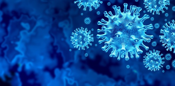
A Microneedle Vaccine Patch Printer For Thermostable MRNA Vaccines

What if you could get vaccinated with the ease of putting on an adhesive bandage? This is the promise of microneedle patches (MNP), which are essentially what they sound like. These would also have uses in diagnostics that might one day obliviate the need for drawing blood. The one major issue with MNPs is their manufacturing, which has been a laborious and highly manual process. In a recent paper in Nature Biotechnology researchers detail the construction and testing of a MNP printer, or microneedle vaccine printer (MVP) that can print dissolving polymers containing stabilized mRNA vaccine.

These mRNA strands are as usual encapsulated in a liquid nanoparticle container, which is mixed with the soluble and biocompatible polymer. This mixture is then added to a mold and dried, after which it retains the microneedle structure of the mold. On tests involving pig skin, the MNPs were capable of penetrating the skin and delivering the vaccine contained in the needles. Produced patches were shown to be shelf-stable for at least six months, which would make these ideal for vaccine distribution in areas where refrigeration and similar are problematic.

Using MNPs for delivering vaccines has previously been researched for e.g. delivering rotavirus and poliovirus vaccine, and a 2021 study in Nature Biomedical Engineering looked at the viability of using MNPs to rapidly sample protein biomarkers in interstitial fluid, which could make diagnostics for certain biomarkers as uncomplicated as putting on the patch, removing it and examining it, removing the need for drawing blood or sampling large amounts of interstitial fluid for external analysis.

If the concept of the MVP and similar MNP printers can be commercialized, it might make it possible to strongly shorten the supply chain for vaccines in less developed regions, while also enabling diagnostics that are very costly and cumbersome today.

USB Borescope Lets Doctors Hone Intubation Skills On The Cheap

One of the most critical skills in emergency medicine is airway management. Without a patent airway, a patient has about four minutes to live, so doctors and paramedics put a huge amount of effort into honing their intubation skills. They have to be able to insert an endotracheal tube quickly and efficiently, without damaging sensitive structures like the vocal cords. It’s a tricky skill to master without a ton of practice.

The perfect tool to practice these skills is a video laryngoscope, but these are wildly expensive and reserved for clinical use. Luckily, with a little ingenuity and a cheap USB borescope, [Dr. Adam Blumenberg] and [Dr. Erin Falk] were able to come up with this low-cost video-assisted laryngoscopy setup to reach as many students as possible. The idea is to use a single-use laryngoscope blade, which replicates the usual tool used to visualize the patient’s vocal cords. The blade is made from clear plastic, which makes it perfect for the application. The borescope is passed through an opening in the blade and affixed to it with adhesives. A little Dremel work might be necessary to get the optical axes of the blade and the camera to line up; failing that, there’s always the option to disassemble the camera to get a better angle.

The chief advantage of this setup, aside from being cheap, is that it’s something that it’s not intended to be used on patients. Along with an airway manikin, the tricked-out borescope can sit in a conference room waiting for students to have a go. Using a large screen allows the whole group to watch the delicate procedure and learn from the mistakes of others. It may not be as detailed a simulation environment as some, but “blade time” is really what counts here.

Insulin Pump Teardown Shows One Motor Does Many Jobs

Modern insulin pumps are self-contained devices that attach to a user’s skin via an adhesive patch, and are responsible for administering insulin as needed. Curious as to what was inside, [Ido Roseman] tore down an Omnipod Dash and took some pictures showing what was inside.

A single motor handles inserting the cannula into the skin, retracting the insertion needle, and administering insulin.

These devices do quite a few things. In addition to holding a reservoir of insulin, they automatically insert a small cannula (thin tube) through the skin after being attached, communicate wirelessly with a control system, and pump insulin through the cannula as needed. All in a sealed and waterproof device. They are also essentially disposable, so [Ido] was curious about what kind of engineering went into such a thing.

The teardown stops short of identifying exactly how all the mechanisms inside work, but [Ido] was able to learn a few interesting things. For example, all of the mechanical functions — inserting the cannula with the help of a needle (and retracting the needle afterwards) and pumping insulin — are all accomplished by one motor and some clever mechanical engineering.

The electronics consist of a PCB with an NXP EX2105F 32-bit Arm7 microcontroller, a second chip that is likely responsible for the wireless communications, three captive LR44 button cells, and hardly a passive component in sight.

The software and communications side of an insulin pump like this one has had its RF communications reverse-engineered with the help of an SDR, a task that took a lot more work than one might expect. Be sure to follow that link if you’re interested in what it can take to get to the bottom of mystery 433 MHz communications on a device that isn’t interested in sharing.

Soft Robotic System For In Situ 3D Bioprinting And Endoscopic Surgery

The progress of medical science has meant increasingly more sophisticated ways to inspect and repair the body, with a shift towards ever less invasive and more effective technologies. An exciting new field is that of in situ tissue replacement in a patient, which can be singular cells or even 3D printed tissues. This in vitro approach of culturing replacement tissues comes however with its share of issues, such as the need for a bioreactor. A more straightforward approach is printing the cells in vivo, meaning directly inside the patient’s body, as demonstrated by a team at the University of New South Wales Sydney with a soft robot that can print layers of living cells inside for example a GI tract.

In their paper, the team — led by [Dr Thanh Nho Do] and PhD student [Mai Thanh Thai] — describe the soft robot that is akin to a standard endoscope, but with a special head that has four soft microtubule artificial muscles (SMAM) for three degrees of freedom and fabric bellow actuators (FBA) that provide the motion desired by the remote controller. The system is configured in such a way that the operator inputs the rough intended motions, which are then smoothed by the software before the hydraulics actuate the head.

In a test on a simulated GI tract, the researchers were able to manipulate a prototype, and deposit a range of materials from the installed syringes. They envision that a system like this could be used as with endoscopes and laparoscopy to not only accurately deposit replacement cells inside the patient’s body, but also to perform a range of other surgical interventions, whereby the surgeon is supported by the system’s software, rather than manipulating the instruments directly.

3D Printed Artificial Nose Is Totally Vegan

Prosthetics are complicated, highly personal things. They must often be crafted and customized precisely to suit the individual. Additive manufacturing is proving a useful tool in this arena, as demonstrated by a new 3D printed nose design developed at Swansea University. And a bonus? It’s vegan, too!

Often, cartilage from the ribcage is used when reconstructing a patient’s nose. However, this procedure is invasive and can lead to health complications. Instead, a nanocellulose hydrogel made from pulped softwood, combined with hyaluronic acid, may be a viable printable material for creating a scaffold for cartilage cells. The patients own cartilage cells can be used to populate the scaffold, essentially growing a new nose structure from scratch. The technique won’t just be limited to nose reconstructions, either. It could also help to recreate other cartilage-based structures, such as the ear.

As with all new medical technologies, the road ahead is long. Prime concerns involve whether the material is properly bio-compatible, particularly where the immune system is concerned. However, the basic idea is one that’s being pursued in earnest by researchers around the world, whether for cosmetic purposes or to grow entire organs. As always, if you’re secretly 3D printing functional gallbladders in your basement, don’t hesitate to drop us a line.

How Tattoos Interact With The Immune System Could Have Impacts For Vaccines

Tattoos are an interesting technology. They’re a way of marking patterns and designs on the skin that can last for years or decades. All this, despite the fact that our skin sloughs off on a regular basis!

As it turns out, tattoos actually have a deep and complex interaction with our immune system, which hold some of the secrets regarding their longevity. New research has unveiled more insight into how the body responds when we get inked up.

Continue reading “How Tattoos Interact With The Immune System Could Have Impacts For Vaccines”

A clear flexible PCB with a number of gold electrodes on one end. It is wrapped over a black cable to demonstrate its flexibility. A set of dashed white lines goes from one end to a zoomed in image of the circuit structure inset in the top right of the image.

Biohybrid Implant Patches Broken Nerves With Stem Cells

Neural interfaces have made great strides in recent years, but still suffer from poor longevity and resolution. Researchers at the University of Cambridge have developed a biohybrid implant to improve the situation.

As we’ve seen before, interfacing electronics and biological systems is no simple feat. Bodies tend to reject foreign objects, and transplanted nerves can have difficulty assuming new roles. By combining flexible electronics and induced pluripotent stem cells into a single device, the researchers were able to develop a high resolution neural interface that can selectively bind to different neuron types which may allow for better separation of sensation and motor signals in future prostheses.

As is typically the case with new research, the only patients to benefit so far are rats and only on the timescale of the study (28 days). That said, this is a promising step forward for regenerative neurology.

We’re no strangers to bioengineering here. Checkout how you can heal faster with electronic bandages or build a DIY vibrotactile stimulator for Coordinated Reset Stimulation (CRS).

(via Interesting Engineering)