Most posts here are electrical or mechanical, with a few scattered hacks from other fields. Those who also keep up with advances in biomedical research may have noticed certain areas are starting to parallel the electronics we know. [Dr. Rajib Shubert] is in one such field, and picked up on the commonality as well. He thought it’d be interesting to bridge the two worlds by explaining his research using analogies familiar to the Hackaday audience. (Video also embedded below.)
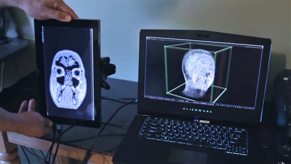
He laid the foundation with a little background, establishing that we’ve been able to see individual static neurons for a while via microscope slides and such, and we’ve been able to see activity of the whole living brain via functional MRI. These methods gradually improved our understanding of neurons, and advances within the past few years have reached an intersection of those two points: [Dr. Shubert] and colleagues now have tools to peer inside a functional brain, teasing out how it works one neuron at a time.
[Dr. Shubert]’s talk makes analogies to electronics hardware, but we can also make a software analogy treating the brain as a highly optimized (and/or obfuscated) piece of code. Virus stamping a single cell under this analogy is like isolating a single function, seeing who calls it, and who it calls. This pairs well with optogenetics techniques, which can be seen as like modifying a function to see how it affects results in real time. It certainly puts a different meaning on the phrase “working with live code”!
Continue reading “Reverse-Engineering Brains, One Neuron At A Time”


 As Director of Apollo Flight Computer Programming,
  Physicist