Brain on a chip setup with a hand and a dropper

Gray Matter On A Chip: Building An Artificial Brain With Luminol

Ever wondered if you could build a robot controlled by chemical reactions? [Marb] explores this wild concept in his video, merging chemistry and robotics in a way that feels straight out of sci-fi. From glowing luminol reactions to creating artificial logic gates, [Marb]—a self-proclaimed tinkerer—takes us step-by-step through crafting the building blocks for what might be the simplest form of a chemical brain.

In this video, the possibilities of an artificial chemical brain take centre stage. It starts with chemical reactions, including a fascinating luminol-based clock reaction that acts as a timer. Then, a bionic robot hand makes its debut, complete with a customised interface bridging the chemical and robotic worlds. The highlight? Watching that robotic hand respond to chemical reactions!

The project relies on a “lab-on-a-chip” approach, where microfluidics streamline the processes. Luminol isn’t just for forensic TV shows anymore—it’s the star of this experiment, with resources like this detailed explanation breaking down the chemistry. For further reading, New Scientist has you covered.

We’ve had interesting articles on mapping the human brain before, one on how exactly brains might work, or even the design of a tiny robot brain. Food for thought, or in other words: stirring the gray matter.

Continue reading “Gray Matter On A Chip: Building An Artificial Brain With Luminol”

Print Your Own Brain Lamp From MRI Data

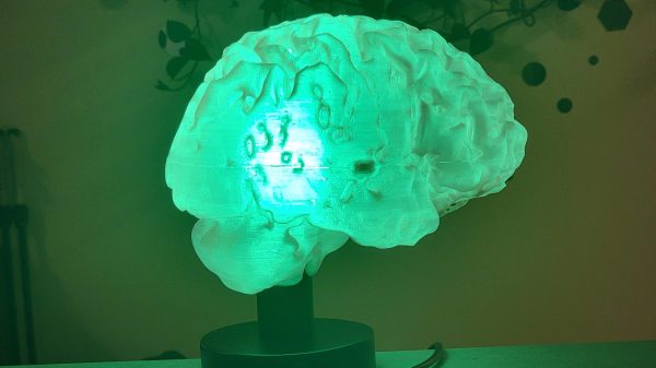
MRIs generally fall somewhere on the scale from boring to stressful depending on why you’re having one and how claustrophobic you get. Regardless, they’re a wonderful diagnostic tool and they’ve saved thousands if not millions of lives over the years. In a fun use of the technology, [mandalaFractals] has shown us how to make a 3D-printed brain lamp using an MRI scan of the head.

The build starts with an off-the-shelf lamp base and a smart LED bulb as the light source, though you could swap those out as desired for something like a microcontroller, a USB power supply, and addressable LEDs if you were so inclined. The software package Slicer is then used to take an MRI brain scan and turn it into something that you can actually 3D print. It’ll take some cleaning up to remove artifacts and hollow it out, but it’s straightforward enough to get a decent brain model out of the data. Alternatively, you can use someone else’s if you don’t have your own scan. Then, all you have to do is print it in a couple of halves, and pop it on the lamp base, and you’re done!

It’s a pretty neat build. Who wouldn’t love telling their friends that their new brain lamp was an accurate representation of their own grey noodles, after all? It could be a fun gift next time Halloween rolls around, too!

Meanwhile, if you’ve got your own MRI hacks that you’ve been cooking up, don’t hesitate to let us know!

Microbubbles And Ultrasound: Getting Drugs Through The Blood-Brain Barrier

The brain is a rather important organ, and as such, nature has gone to great lengths to protect it. The skull provides physical protection against knocks and bumps, but there’s a lesser-known defense mechanism at work too: the blood-brain barrier. It’s responsible for keeping all the nasty stuff – like bacteria, viruses, and weird chemicals – from messing up your head.

The blood-brain barrier effectively acts as a filter between the body’s circulatory system and the brain. However, it also frustrates efforts to deliver drugs directly to the brain for treating conditions like brain tumors. Now, scientists have developed a new technique that may allow critical life saving drugs to get through the barrier with the help of ultrasound technology. 

Continue reading “Microbubbles And Ultrasound: Getting Drugs Through The Blood-Brain Barrier”

MRI Resolution Progresses From Millimeters To Microns

Neuroscientists have been mapping and recreating the nervous systems and brains of various animals since the microscope was invented, and have even been able to map out entire brain structures thanks to other imaging techniques with perhaps the most famous example being the 302-neuron brain of a roundworm. Studies like these advanced neuroscience considerably but even better imaging technology is needed to study more advanced neural structures like those found in a mouse or human, and this advanced MRI machine may be just the thing to help gain better understandings of these structures.

A research team led by Duke University developed this new MRI technology using an incredibly powerful 9.4 Tesla magnet and specialized gradient coils, leading to an image resolution an impressive six orders of magnitude higher than a typical MRI. The voxels in the image measure at only 5 microns compared to the millimeter-level resolution available on modern MRI machines, which can reveal microscopic details within brain tissues that were previously unattainable. This breakthrough in MRI resolution has the potential to significantly advance understanding of the neural networks found in humans by first studying neural structures in mice at this unprecedented detail.

The researchers are hopeful that this higher-powered MRI microscope will lead to new insights and translate directly into advancements healthcare, and presuming that it can be replicated, used on humans safely, and becomes affordable, we would expect it to find its way into medical centers as soon as possible. Not only that, but research into neuroscience has plenty of applications outside of healthcare too, like the aforementioned 302-neuron brain of the Caenorhabditis elegans roundworm which has been put to work in various robotics platforms to great effect.

Continue reading “MRI Resolution Progresses From Millimeters To Microns”

A clear droplet sits on a blue PCB with gold traces. A syringe with a drop of clear liquid sits above the droplet.

Grow Your Own Brain Electrodes

Bioelectronics has been making great strides in recent years, but interfacing rigid electrical components with biological systems that are anything but can prove tricky. Researchers at the Laboratory for Organic Electronics (LOE) have found a way to bridge the gap with conductive gels. (via Linköping University)

Outside the body, these gels are non-conductive, but when injected into a living animal, the combination of gel and the body’s metabolites creates a conductive electrode that can move with the tissue. This is accompanied by a nifty change in color which makes it easy for researchers to see if the electrode has formed properly.

Side-by-side images of a zebrafish tail. Both say "Injected gel with LOx:HRP" at the top with an arrow going to the upper part of the tail structure. The left says "t=0 min" and "Injected with gel GOx:HRP" along the bottom with an arrow going to the lower part of the tail structure. The tail shows darkening in the later image due to formation of bioelectrodes.

Applications for the technology include better biological sensors and enhanced capabilities for future brain-controlled interfaces. The study was done on zebrafish and medicinal leeches, so it will be awhile before you can pick up a syringe of this stuff at your local computer store, but it still offers a tantalizing glimpse of the future.

We’ve covered a few different brain electrodes here before including MIT’s 3D printed version and stentrodes.

Auditory Brainstem Implants: The Other Bionic Hearing Device

You might have heard of the cochlear implant. It’s an electronic device also referred to as a neuroprosthesis, serving as a bionic replacement for the human ear. These implants have brought an improved sense of hearing to hundreds of thousands around the world.

However, the cochlear implant isn’t the only game in town. The auditory brain stem implant is another device that promises to bring a sense of sound to those without it, albeit by a different route.

Continue reading “Auditory Brainstem Implants: The Other Bionic Hearing Device”

The Real Science (Not Armchair Science) Of Consciousness

Among brain researchers there’s a truism that says the reason people underestimate how much unconscious processing goes on in your brain is because you’re not conscious of it. And while there is a lot of unconscious processing, the truism also points out a duality: your brain does both processing that leads to consciousness and processing that does not. As you’ll see below, this duality has opened up a scientific approach to studying consciousness.

Are Subjective Results Scientific?

Researcher checking fMRI images.
Checking fMRI images.

In science we’re used to empirical test results, measurements made in a way that are verifiable, a reading from a calibrated meter where that reading can be made again and again by different people. But what if all you have to go on is what a person says they are experiencing, a subjective observation? That doesn’t sound very scientific.

That lack of non-subjective evidence is a big part of what stalled scientific research into consciousness for many years. But consciousness is unique. While we have measuring tools for observing brain activity, how do you know whether that activity is contributing to a conscious experience or is unconscious? The only way is to ask the person whose brain you’re measuring. Are they conscious of an image being presented to them? If not, then it’s being processed unconsciously. You have to ask them, and their response is, naturally, subjective.

Skepticism about subjective results along with a lack of tools, held back scientific research into consciousness for many years. It was taboo to even use the C-word until the 1980s when researchers decided that subjective results were okay. Since then, here’s been a great deal of scientific research into consciousness and this then is a sampling of that research. And as you’ll see, it’s even saved a life or two.

Continue reading “The Real Science (Not Armchair Science) Of Consciousness”