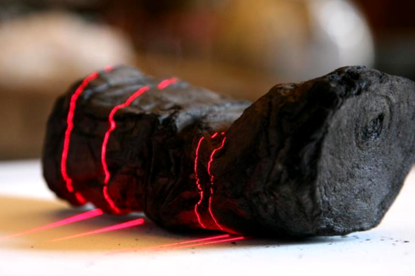
Imagine trying to read a 2000-year old scroll from an ancient civilization. Now imagine that scroll is rolled up, and in a delicate, charred, carbonized form, having been engulfed by the fiery eruption of a volcano. The task would seem virtually impossible, and the information in the scroll lost forever. Right?|
We’ve recently got a look at how [Ken Shirriff] used an industrial CT scanner as a reverse engineering tool. The results were spectacular, with pictures that clearly showed the internal arrangement of parts that haven’t seen the light of day since the module was potted back in the 60s. And now, [Ken]’s cohort [Curious Marc] has dropped a video with more detail on the wonderful machine, plus deep dives into more Apollo-era hardware
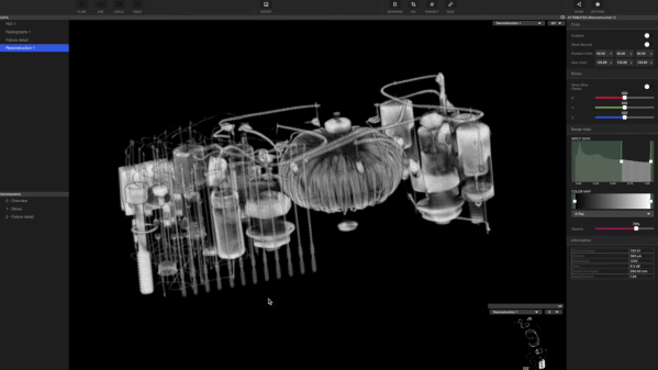
If you liked seeing the stills [Ken] used to reverse engineer the obscure flip-flop module, you’re going to love seeing [Marc] using the Lumafield scanner’s 3D software to non-destructively examine several Apollo artifacts. First to enter the sample chamber of the CT scanner was a sealed module called the Central Timing Equipment, which served as the master clock for the Apollo Command Module. The box’s magnesium case proved to be no barrier to the CT scanner’s beam, and the 3D model that was built up from a series of 2D images was astonishingly detailed. The best part about the virtual models is the ability to slice through them in any plane — [Marc] used this feature to hunt down the clock’s quartz crystal. Continue reading “A Deeper Dive Into Reverse Engineering With A CT Scanner”→
[Pyrotechnical] thought about buying a CAT scanner and found out they cost millions of dollars. So he decided to build one for about $200 using a salvage X-ray tube and some other miscellaneous parts. A scintillating detector provides the image for pick up with a camera phone. The control? An Arduino, what else? You can watch the video below, but due to plenty of NSFW language, you might want to put your headphones on if you don’t want to shock anyone.
Of course, you need to be careful when working with energetic X-rays. To keep away from the X-ray source, [Pyrotechnical] used a Roku remote and an IR sensor to control the device from afar. The electronics is pretty easy. You just have to rotate a turntable and trigger the camera while lighting up the X-ray tube.
It’s that time of year again — the 2022 Hackaday Prize has officially launched, and we’re excited to see what it turns out. This year’s theme is “Sustainability, Resilience, and Circularity,” and just in time, too; if the last couple of years has taught us anything, it’s that we’ve got a lot of failure points built into the systems that run our world. As broken as things are, it’s tempting to just curl up in a ball and pretend everything’s fine, but that’s not how hackers respond to adversity. We need to control what we can control, and there’s plenty of work to be done. From sustainable energy ideas to ways to reduce the amount of stuff we throw away, from breathing new life into old equipment to building communities that can take care of themselves, there’s plenty of work to be done. So get over to the Hackaday Prize page, check out the launch summit video if you need some inspiration, and get hacking. And hurry up — things are only going to get better if people like us make it happen.
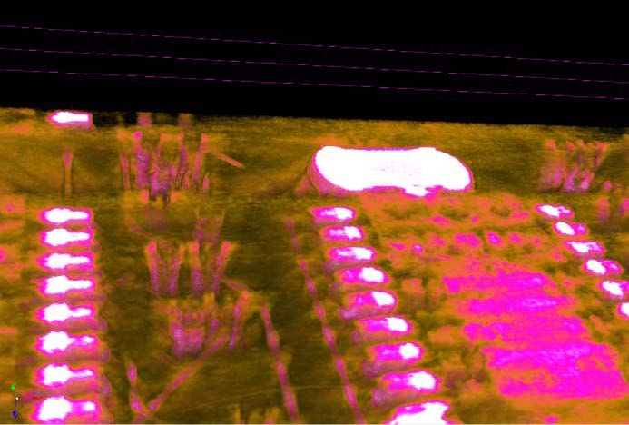
At risk of getting any ASMR buffs who might be reading cranky because there’s no audio, [Chris], or [@no1089] on Twitter, has gifted us with this visually stunning scan of his Maxim MAX86160 in-ear heart monitor mounted on a rigidflex PCB. You can take a look, in the video below the break.
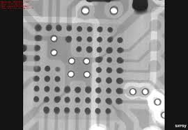
If you’re wondering why anyone would scan a board, other than boredom, know that it’s actually quite common. X-Ray machines are commonly used as a quick, passive way to check a board that’s fresh off the production line. These aren’t the X-Rays like those of broken bones you’re (hopefully not too) used to seeing though, they’re Computed Tomography scans (CT scans, CAT scans), in effect just 3D X-Rays.
For electronics manufacturers and assemblers, CT scans are incredibly useful because they provide a non-destructive way to check for errors. For example, how do you know if that middle BGA pin is actually soldered correctly? You could run a functional test and make sure everything is working (at least, everything you check), but that takes time. The longer it takes to validate, the higher the manufacturing cost. In manager speak: “cost bad. Fast good.”
It’s also common to use a CT scan to create a full 3D model of a board. This makes it easy to check every little detail, especially the ones that are visually obscured by surface mount devices or critical signal paths that are buried under board layers.
Highlight of solder joints on small-outline integrated circuit (SOIC) to a PCB’s pads.
But we know you really want more of this video, but better. And we’ve got the goods. For the chill folk among you, here’s a 55-minute version without all the CT scan info cluttering the screen. For those of you currently blasting eDM in your headphones, here’s a 30 second clip of it looping at ~5x speed. Eat your heart out:
Fran Piernas likes to push the envelope a bit with projects that others might shy away from. A quick glance at his Hackaday.io profile reveals a few of the exciting projects he’s been working on recently, including a DIY X-ray machine and the high-voltage driver needed to run it. Not only that, he’s recently taken his home-brew X-ray rig to the next level – a computed tomography (CT) scanner. His YouTube channel also has some exciting stuff using potentially lethal voltages and ionizing radiation.
Please join us for this Hack Chat, in which we’ll cover:
How one safely works with high voltage and ionizing radiation;
Sourcing uncommon components like X-ray tubes;
How Fran decided to start playing at the edge of the danger zone; and
What sort of experiments he has in mind for the future.
You are, of course, encouraged to add your own questions to the discussion. You can do that by leaving a comment on the X-rays and high-voltage Hack Chat and we’ll put that in the queue for the Hack Chat discussion.
Click that speech bubble to the right, and you’ll be taken directly to the Hack Chat group on Hackaday.io. You don’t have to wait until Wednesday; join whenever you want and you can see what the community is talking about.
The 3D printers we’re most familiar with use the fused deposition process, in which hot plastic is squirted out of a nozzle, to build up parts on a layer by layer basis. We’ve also seen stereolithography printers, such as the Form 2, which use a projector and a special resin to produce parts, again in a layer-by-layer method. However, a team from the University of North Carolina were inspired by CT scanners, and came up with a novel method for producing 3D printed parts.
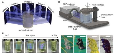
The process, as outlined in the team’s paper.
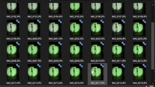
The technique is known as Computed Axial Lithography. The team describe the system as working like a CT scan in reverse. The 3D model geometry is created, and then a series of 2D images are created by rotating the part about the vertical axis. These 2D images are then projected into a cylindrical container of photosensitive resin, which rotates during the process. Rather than building the part out of a series of layers in the Z-axis, instead the part is built from a series of axial slices as the cylinder rotates.
The parts produced have the benefit of a smooth surface finish and are remarkably transparent. The team printed a variety of test objects, including a replica of the famous Thinker sculpture, as well as a replica of a human jaw. Particularly interesting is the capability to make prints which enclose existing objects, demonstrated with a screwdriver handle enclosing the existing steel shank.
It’s a technique which could likely be reproduced by resourceful makers, assuming the correct resin isn’t too hard to come by. The resin market is hotting up, with Prusa announcing new products at a recent Makerfaire. We’re excited to see what comes next, particularly as the high cost of resin is reduced by economies of scale. Video after the break.