Video: Bil Herds Looks At Mitosis

I loved my science courses when I was in Junior High School; we leaned to make batteries, how molecules combine to form the world we see around us, and basically I got a picture of where we stood in the  scheme of things, though Quarks had yet to be discovered at the time.

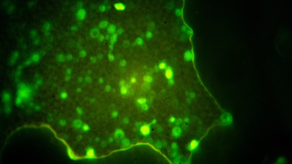
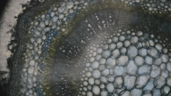
In talking with my son I found out that there wasn’t much budget for Science learning materials in his school system like we had back in my day, he had done very little practical hands-on experiments that I remember so fondly. One of those experiments was to look and draw the stages of mitosis as seen under a Microscope. This was amazing to me back in the day, and cemented the wonder of seeing cell division into my memory to this day, much like when I saw the shadow of one of Jupiter’s moons with my own eyes!

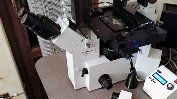
Now I have to stop and tell you that I am not normal, or at least was not considered to be a typical young’un growing up near a river in rural Indiana in the 60’s. I had my own microscope; it quite simply was my pride and joy. I had gotten it while I was in the first or second grade as a present and I loved the thing. It was just horrible to use in its later years as lens displaced, the focus rack became looser if that was possible, and dirt accumulated on the internal lens; and yet I loved it and still have it to this day! As I write this, I realize that it’s the oldest thing that I own. (that and the book that came with it).

Today we have better tools and they’re pretty easy to come by. I want to encourage you to do some science with them. (Don’t just look at your solder joints!) Check out the video about seeing mitosis of onion cells through the microscope, then join me below for more on the topic!

Continue reading “Video: Bil Herds Looks At Mitosis”

The Smallest Cell Phone Picture

Mobile phones are the photography tool for most of us, but they are a blunt tool. If you love astrophotography, you buy a DSLR and a lens adapter. Infrared photography needs camera surgery or a special unit. If you want to look closer to home, you may have a microscope with a CCD. Your pocket computer is not manufactured for microscopy, but that does not mean it cannot be convinced. Most of us have held our lens up to the eyepiece of some binoculars or a microscope, and it sort of works, but it is far from perfect. [Benedict Diederich] and a team are proving that we can get darn beautiful images with a microscope, a phone holder, and some purpose-built software on an Android phone with their cellSTORM.

The trick to getting useful images is to compare a series of pictures and figure out which pixels matter and which ones are noisy. Imagine someone shows you grainy nighttime footage from an outdoor security camera. When you pause, it looks like hot garbage, and you can’t tell the difference between a patio chair and a shrubbery. As it plays, the noisy pixels bounce around, and you figure out you’re looking at a spruce bush, and that is roughly how the software parses out a crisp image. At the cost of frame rate, you get clarity, which is why you need a phone holder. Some of their tests took minutes, so astrophotography might not fare as well.

We love high-resolution pictures of tiny things and that isn’t going to change anytime soon.

Thank you [Dr. Nicolás De Francesco] for the tip.

Focus Stacking For Tiny Subjects

Focus stacking is a photographic technique in which multiple exposures are taken of a subject, with the focus distance set to different lengths. These images are then composited together to create a final image with a greater depth of field than is possible with a single exposure. [Peter Lin] built a rig for accurate focus stacking with very small subjects.

The heart of the rig is a motion platform consisting of a tiny stepper motor fitted with a linear slide screw. This is connected to an Arduino or PIC with a basic stepper driver board. While the motor does not respond well to microstepping or other advanced techniques, simply driving it properly can give a resolution of 15 μm per step.

The motor/slide combination is not particularly powerful, and thus cannot readily be used to move the camera or optics. Instead, the rig is designed for photography of very small objects, in which the rail will move the subject itself.

It’s a tidy build that would serve well for anyone regularly doing macro focus stack photography. If you’ve been trying to better photograph your insect collection, this one is for you. It’s a valuable technique and one that applies to microscopy too. Video after the break.

Continue reading “Focus Stacking For Tiny Subjects”

Seeing Transistors Switch In Infrared

In the hacker and DIY community, there are people who have exceptional knowledge and fantastic tools. These people are able to do what others could only dream about, and that others can only browse eBay looking for that one tool they need to do the job. One of these such people is [John McMaster]. He is the resident expert on looking inside integrated circuits. He drops acid on a chip, and he can tell you exactly how it works on the inside.

At the hardwear.io conference, [John] shared one of his techniques for reverse-engineering intgrated circuits. He’s doing this by simply looking at the transistors, and looking at the light they give off. He’s also looking at the wrong side of the die.

The technique [John] is using is properly called backside analysis, or looking at the infrared emissions of electron recombinations. This happens at the junction of every transistor when it’s active, and these photons are emitted at the bandgap of silicon, or about 1088 nm, far into the infrared. This sort of thing has been done before by [nedos] at CCC in 2013, but rarely have we seen a deep dive into the tools and techniques needed to look at the reverse side of an IC and see the photons coming off.

An IC, seen in infrared

There are several tools [John] used for this work, and he actually did a good comparison of different camera technologies used to image infrared photon emissions from integrated circuits. InGaAs cameras are expensive, but they offer high sensitivity. New back-illuminated CMOS cameras and cooled CCDs normally reserved for astrophotography were also tested, and as always, you get what you pay for; the most expensive cameras worked best, but there were ways you could make the cheap ones work.

As with any camera work, preparing the lighting is of utmost importance. This includes an IR pass filter, and using only LED lighting in the lab with no sunlight, incandescent, or halogen light bulbs in the room — you don’t want any IR, after all. A NIR objective in the microscope was sourced from eBay, for about 1/10th the normal cost, because the objective had a small, insignificant scratch. Using this NIR objective made the image twice as bright as any other method. You can successfully image a chip with this, and [John] tested the setup on a resistor inside a CD4050 chip; the resistor glowed a slight purple, the color you would expect with infrared sensors. But can it work with I/O levels in a more modern chip? Also, yes. It needs some Photoshop to process, and stretching the 12-bit or 16-bit color space into an 8-bit color space, but it does work.

Finally, the supreme achievement of doing backside IR analysis. Is that possible with even this minimal setup? This requires some preparation; the silicon substrate in an IC is transparent in IR, but there is attenuation and this is especially important when the substrate is 300 um thick. This needs to be shaved down to about 25 um thick, which surprisingly is best done with fine sandpaper and a finger.

While few IR emissions were observed via backside emissions, the original plan wasn’t to completely analyze the chip, but merely to do some floor planning. For this, it worked. It’s a remarkable amount of work to see the inside of a silicon chip.

Fluorescence Microscope On A Hacker’s Budget

Some of biology’s most visually striking images come from fluorescence microscopes. Their brilliant colors on black look like a neon sign from an empty highway. A brand new fluorescence microscope is beyond a hacker’s budget and even beyond some labs’, but there are ways to upgrade an entry-level scope for the cost of a few cups of coffee. [Justin Atkin] of The Thought Emporium published a scope hacking video which can also be seen below. He is becoming a reputed scope modder.

This video assumes a couple of things for the $10 price tag. The first premise is that you already have a scope, a camera adapter, and a camera capable of shooting long exposures. The second premise is that you are willing to break the seals and open the scope to make some reversible mods. Since you are reading Hackaday, maybe that is a given.

The premise is simple compared to the build, which is not rocket surgery, the light source from below illuminates the subject like a raver, and the filter removes any light that isn’t spectacular before it gets to the camera.

Continue reading “Fluorescence Microscope On A Hacker’s Budget”

Plant Biology Is A Gateway

Too many college students have been subject to teachers’ aids who think they are too clever to be stuck teaching mere underclassmen. For that reason, [The Thought Emporium] is important because he approaches learning with gusto and is always ready to learn something new himself and teach anyone who wants to learn. When he released a video about staining and observing plant samples, he avoided the biggest pitfalls often seen in college or high school labs. Instead of calling out the steps by rote, he walks us through them with useful camera angles and close-ups. Rather than just pointing at a bottle and saying, “the blue one,” he tells us what is inside and why it is essential. Instead of telling us precisely what we need to see to get a passing grade, he lets our minds wonder about what we might see and shows us examples that make the experiment seem exciting. The video can also be seen below the break.

The process of staining can be found in a biology textbook, and some people learn best by reading, but we haven’t read a manual that makes a rudimentary lab seem like the wardrobe to Narnia, so he gets credit for that. Admittedly, you have to handle a wicked sharp razor, and the chance of failure is never zero. In fact, he will tell you, the opportunities to fail are everywhere. The road to science isn’t freshly paved, it needs pavers.

If a biology lab isn’t in your personal budget, a hackerspace may have one or need one. If you are wondering where you’ve heard [The Thought Emporium]’s voice before, it is because he is fighting lactose intolerance like a hacker.

Continue reading “Plant Biology Is A Gateway”

Motorized Stage Finesses The Microscopic World

No matter how fine your fine motor skills may be, it’s really hard to manipulate anything on the stage of a microscope with any kind of accuracy. One jitter or caffeine-induced tremor means the feature of interest on the sample you’re looking at shoots off out of the field of view, and getting back to where you were is a tedious matter of trial and error.

Mechanical help on the microscope stage is nice, and electromechanical help is even better, but a DIY fully motorized microscope stage with complete motion control is the way to go for the serious microscopist on a budget. Granted, not too many people are in [fabiorinaldus]’ position of having a swell microscope like the Olympus IX50, and those that do probably work for an outfit that can afford all the bells and whistles. But this home-brew stage ticks off all the boxes on design and execution. The slide is moved across the stage in two dimensions with small NEMA-8 steppers and microstepping controllers connected to two linear drives that are almost completely 3D-printed. The final resolution on the drives is an insane 0.000027344 mm. An Arduino lives in the custom-built control box and a control pad with joystick, buttons, and an OLED display allow the stage to return to set positions of interest. It’s really quite a build.

We’ve featured a lot of microscope hacks before, most of them concerning the reflective inspection scopes we all seem to covet for SMD work. But that doesn’t mean we haven’t shown love for optical scopes before, and electron microscopes have popped up a time or two as well.

Continue reading “Motorized Stage Finesses The Microscopic World”