Turn Medical Imaging From 2D Into 3D With Just $10

One of the modern marvels in our medical toolkit is ultrasound imaging. One of its drawbacks, however, is that it displays 2D images. How expensive do you think it would be to retrofit an ultrasound machine to produce 3D images? Try a $10 chip and pennies worth of plastic.

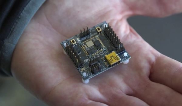
While — of all things — playing the Wii with his son, [Joshua Broder, M.D], an emergency physician and associate professor of surgery at [Duke Health], realized he could port the Wii’s gyroscopic sensor to ultrasound technology. He did just that with the help of [Matt Morgan, Carl Herickhoff and Jeremy Dahl] from [Duke’s Pratt School of Engineering] and [Stanford University]. The team mounted the sensor onto the side of the probe with a 3D printed collar. This relays the orientation data to the computer running software that sutures the images together into a complete 3D image in near real-time, turning a $50,000 ultrasound machine into its $250,000 equivalent.

Continue reading “Turn Medical Imaging From 2D Into 3D With Just $10”

Hyperspectral Imaging – Seeing The Unseeable

They say that a picture is worth a thousand words. But what is a picture exactly? One definition would be a perfect reflection of what we see, like one taken with a basic camera. Our view of the natural world is constrained to a bandwidth of 400 to 700 nanometers within the electromagnetic spectrum, so our cameras produce images within this same bandwidth.

Image via Cosmos Magazine.

For example, if I take a picture of a yellow flower with my phone, the image will look just about how I saw it with my own eyes. But what if we could see the flower from a different part of the electromagnetic spectrum? What if we could see less than 400 nm or greater than 700 nm? A bee, like many other insects, can see in the ultraviolet part of the spectrum which occupies the area below 400 nm. This “yellow” flower looks drastically different to us versus a bee.

In this article, we’re going to explore how images can be produced to show spectral information outside of our limited visual capacity, and take a look at the multi-spectral cameras used to make them.  We’ll find that while it may be true that an image is worth a thousand words, it is also true that an image taken with a hyperspectral camera can be worth hundreds of thousands, if not millions, of useful data points. Continue reading “Hyperspectral Imaging – Seeing The Unseeable”

Digitize Your Room With LIDAR

What’s the best way to image a room? A picture? Hah — don’t be so old-fashioned! You want a LIDAR rig to scan the space and reconstruct it as a 3D point map in your computer.

Hot on the heels of [Saulius Lukse]’s scanning thermometer, he’s replaced the thermal camera on their pan/tilt setup with a time-of-flight (TOF) camera — a Garmin LIDAR — capable of 500 samples per second and end up scanning their room in a mere fifteen minutes. Position data is combined with the ranging information to produce a point cloud using Python. Open that file in a 3D manipulation program and you’ll be treated to a sight like this:

Continue reading “Digitize Your Room With LIDAR”

Build A Rhizotron: Image The World Beneath Your Feet

The world of the subsoil is a fascinating place. Our whole ecosystem depends on its variety of fungus, bacteria and detritivore creatures that break down and decay dead matter and provide the nutrients to sustain plants that bring in the energy from the sun.

It’s easy enough to study what is happening beneath the surface, just reach for a trowel. But of course, that’s an imperfect technique, for it only gives a picture of a world you have destroyed, and then at best only a snapshot.

Fungal hypha growth in action

What if you could image underground, take pictures and video of the decay process and the creatures that are its engine? [Josh Williams] was curious how this could be achieved, so after early experiments with buried webcams proved unimpressive he created the Rhizotron. A flatbed scanner waterproofed for burial with plenty of silicone, and driven by a Raspberry Pi. The result was particularly successful, and though he has lost several scanners to water ingress he has collected some impressive imagery which he has posted on the project’s blog. Below the break we’ve included one of his videos taken with the scanner in a compost bucket, in which you can see decomposition aplenty, mating millipedes, spreading fungal hyphae and much more.

Continue reading “Build A Rhizotron: Image The World Beneath Your Feet”

To See Within: Making Medical X-rays

I was a bit of a lost soul after high school.  I dabbled with electrical engineering for a semester but decided that it wasn’t for me – what I wouldn’t give for a do-over on that one. In my search for a way to make money, I stumbled upon radiologic technology – learning how to take X-rays. I figured it was a good way to combine my interests in medicine, electronics, and photography, so after a two-year course of study I got my Associates Degree, passed my boards, and earned the right to put “R.T.(R) (ARRT)” after my name.

That was about as far as that career went. There are certain realities of being in the health care business, and chief among them is that you really have to like dealing with the patients. I found that I liked the technology much more than the people, so I quickly moved on to bigger and better things. But the love of the technology never went away, so I thought I’d take a look at exactly what it takes to produce medical X-rays, and see how it’s changed from my time in the Radiology Department.

Continue reading “To See Within: Making Medical X-rays”

Hacker Reads Magnetic Stripe Card With Flatbed Scanner

[anfractuosus] has been reading magnetic stripe card… optically!

While hackers routinely read and write stripe cards, this is the first time we’ve reported on optically imaging and decoding data from the magnetic stripe. [anfratuosus] used a magnetic developer which is designed to allow visual inspection of the magnetic stripe. The developer uses micron sized iron particles in a suspension which are dropped onto the stripe. To the particles, the magnetic stripe looks like a series of magnets lined up. Long magnets striperepresent 0s and short magnets 1s. With each bit the orientation of the magnet changes, something like the diagram to the right. The magnetic field is strongest where the poles meet. So the iron particles are attracted to these flux reversal points on the stripe creating a visible pattern . There’s an awesome video of the process in action below.

While magnetic developer was designed for debugging faulty recording systems [anfratuosus] went a step further scanning the “developed” card, and writing a tool to decode the images and extract the card data. [anfratuosus] doesn’t mention any particular application, we love this circuitous hack anyway!

Continue reading “Hacker Reads Magnetic Stripe Card With Flatbed Scanner”

We Should 3D Scan People

In a perfect futuristic world you have pre-emptive 3D scans of your specific anatomy. They’d be useful to compare changes in your body over time, and to have a pristine blueprint to aid in the event of a catastrophe. As with all futuristic worlds there are some problems with actually getting there. The risks may outweigh the rewards, and cost is an issue, but having 3D imaging of a sick body’s anatomy does have some real benefits. Take a journey with me down the rabbit hole of 3D technology and Gray’s Anatomy.

Continue reading “We Should 3D Scan People”